

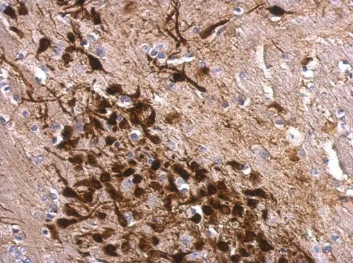
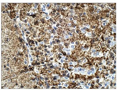
Calretinin antibody detects Calretinin protein at cytosol on mouse fore brain by immunohistochemical analysis. Sample: Paraffin-embedded mouse fore brain. Calretinin antibody (GTX103261) dilution: 1:500.
Antigen Retrieval: Trilogy? (EDTA based, pH 8.0) buffer, 15min
 antibody at 1:250 dilution.
Antigen Retrieval: Trilogy? (EDTA based, pH 8.0) buffer, 15min")
 dilution: 1:5000 The HRP-conjugated anti-rabbit IgG antibody (GTX213110-01) was used to detect the primary antibody.")
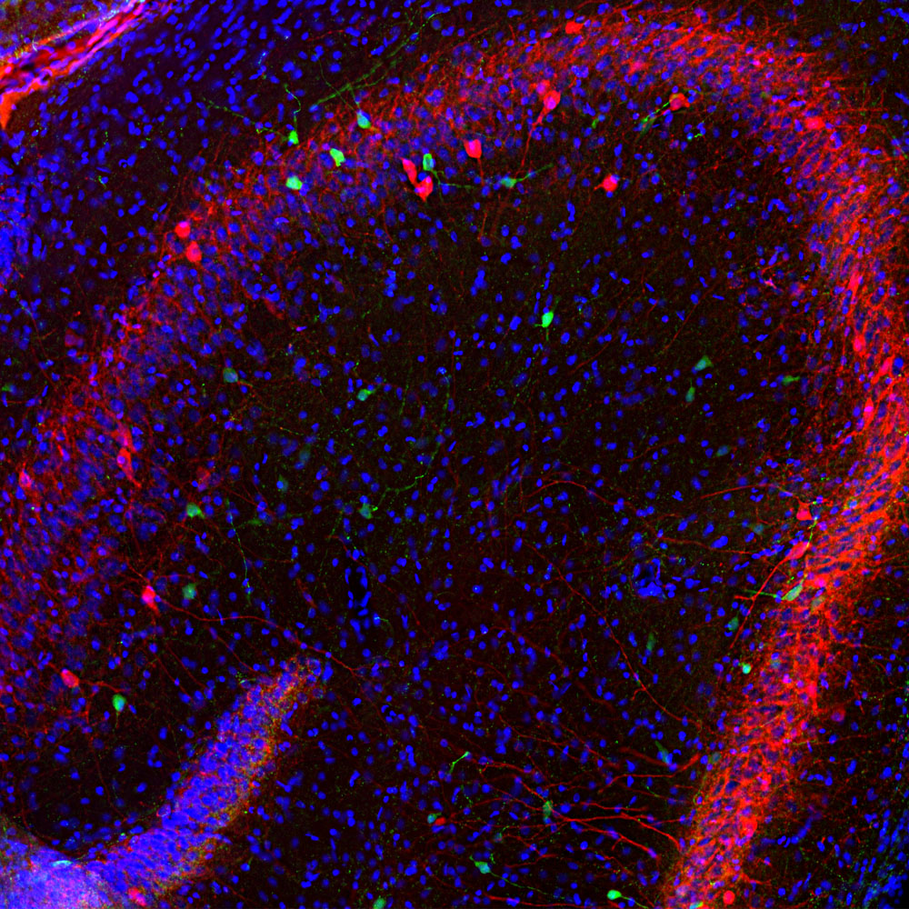
![Calretinin antibody detects Calretinin protein by immunohistochemical analysis. Samples: Frozen Section adult mouse hippocampus. Green: Calretinin protein stained by Calretinin antibody (GTX103261) diluted at 1:250. Red: NF-H, stained by NF-H antibody [GT114] (GTX634289) diluted at 1:500. Blue: Fluoroshield with DAPI (GTX30920).](https://www.genetex.com/upload/website/prouct_img/normal/GTX103261/GTX103261_39876_20171017_IHC-Fr_M_w_23060119_944.webp "Calretinin antibody detects Calretinin protein by immunohistochemical analysis. Samples: Frozen Section adult mouse hippocampus. Green: Calretinin protein stained by Calretinin antibody (GTX103261) diluted at 1:250. Red: NF-H, stained by NF-H antibody [GT114] (GTX634289) diluted at 1:500. Blue: Fluoroshield with DAPI (GTX30920).")
 diluted at 1:200. Blue: Hoechst 33342 staining.")
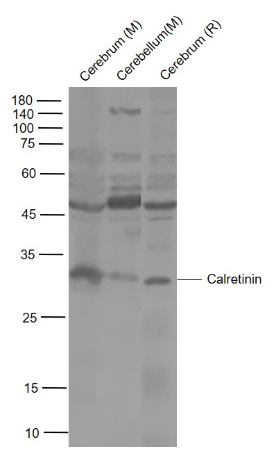
 was separated by 12% SDS-PAGE, and the membrane was blotted with Calretinin antibody (GTX103261) diluted at 1:1000. The HRP-conjugated anti-rabbit IgG antibody (GTX213110-01) was used to detect the primary antibody.")
 A: mouse brain 12% SDS PAGE GTX103261 diluted at 1:1000 The HRP-conjugated anti-rabbit IgG antibody (GTX213110-01) was used to detect the primary antibody.")
 diluted at 1:250. Red: Protein kinase C alpha staining. Blue: Fluoroshield with DAPI (GTX30920).")
 diluted at 1:500. Antigen Retrieval: Citrate buffer, pH 6.0, 15 min")
 were separated by 12% SDS-PAGE, and the membrane was blotted with Calretinin antibody (GTX103261) diluted at 1:1000. The HRP-conjugated anti-rabbit IgG antibody (GTX213110-01) was used to detect the primary antibody. Corresponding RNA expression data for the same cell lines are based on Human Protein Atlas program.")
Calretinin antibody detects Calretinin protein at cytosol on mouse fore brain by immunohistochemical analysis. Sample: Paraffin-embedded mouse fore brain. Calretinin antibody (GTX103261) dilution: 1:500.
Antigen Retrieval: Trilogy? (EDTA based, pH 8.0) buffer, 15min
Calretinin antibody
GTX103261
ApplicationsImmunoFluorescence, Western Blot, ImmunoCytoChemistry, ImmunoHistoChemistry, ImmunoHistoChemistry Frozen, ImmunoHistoChemistry Paraffin
Product group Antibodies
ReactivityHuman, Mouse, Rat
TargetCALB2
Overview
- SupplierGeneTex
- Product NameCalretinin antibody
- Delivery Days Customer9
- Application Supplier NoteWB: 1:500-1:10000. ICC/IF: 1:100-1:1000. IHC-P: 1:100-1:1000. IHC-Fr: 1:100-1:1000. *Optimal dilutions/concentrations should be determined by the researcher.Not tested in other applications.
- ApplicationsImmunoFluorescence, Western Blot, ImmunoCytoChemistry, ImmunoHistoChemistry, ImmunoHistoChemistry Frozen, ImmunoHistoChemistry Paraffin
- CertificationResearch Use Only
- ClonalityPolyclonal
- Concentration0.64 mg/ml
- ConjugateUnconjugated
- Gene ID794
- Target nameCALB2
- Target descriptioncalbindin 2
- Target synonymsCAB29, CAL2, CR, calretinin, 29 kDa calbindin, calbindin 2, (29kD, calretinin), calbindin D29K, testicular secretory protein Li 8
- HostRabbit
- IsotypeIgG
- Protein IDP22676
- Protein NameCalretinin
- Scientific DescriptionThis gene encodes an intracellular calcium-binding protein belonging to the troponin C superfamily. Members of this protein family have six EF-hand domains which bind calcium. Three alternatively spliced transcript variants that encode different proteins have been described. [provided by RefSeq]
- ReactivityHuman, Mouse, Rat
- Storage Instruction-20°C or -80°C,2°C to 8°C
- UNSPSC41116161
Datasheet
Related products
Product group Antibodies
Anti-Calretinin AntibodyA104312
ApplicationsImmunoFluorescence, Western Blot, ImmunoCytoChemistry, ImmunoHistoChemistry
ReactivityBovine, Equine, Human, Mouse, Porcine, Rat
- SizePrice

Product group Antibodies
Anti-Calretinin/CALB2 Antibody Picoband(r)A04255-CARRIER-FREE
ApplicationsFlow Cytometry, ImmunoFluorescence, Western Blot, ELISA, ImmunoCytoChemistry, ImmunoHistoChemistry
ReactivityHuman, Mouse, Rat
TargetCALB2
- SizePrice
Product group Antibodies
Anti-Calretinin Antibody130-10089
ApplicationsELISA
ReactivityHuman
TargetCALB2
- SizePrice

Product group Antibodies
Anti-CALB2 AntibodyAMAB91812
ApplicationsWestern Blot, ImmunoHistoChemistry
ReactivityHuman, Mouse
TargetCALB2
- SizePrice
Product group Antibodies
References
CALB2 Polyclonal AntibodyBS-0062R
ApplicationsImmunoFluorescence, Western Blot, ELISA, ImmunoCytoChemistry, ImmunoHistoChemistry, ImmunoHistoChemistry Frozen, ImmunoHistoChemistry Paraffin
ReactivityBovine, Canine, Equine, Human, Mouse, Porcine, Rat
TargetCALB2
- SizePrice
Product group Antibodies

ApplicationsWestern Blot, ImmunoHistoChemistry, ImmunoHistoChemistry Frozen, ImmunoHistoChemistry Paraffin
ReactivityHuman, Rat
TargetCALB2
- SizePrice
Product group Antibodies
CALB2 antibodyCSB-PA004433
ApplicationsWestern Blot, ELISA, ImmunoHistoChemistry
ReactivityHuman
TargetCALB2
- SizePrice
Product group Antibodies
ApplicationsImmunoPrecipitation, Western Blot, ImmunoCytoChemistry, ImmunoHistoChemistry
ReactivityMouse, Porcine, Rat
TargetCALB2
- SizePrice
Product group Antibodies
Calretinin antibodyGTX14689
ApplicationsImmunoFluorescence, ImmunoPrecipitation, Western Blot, ELISA, ImmunoCytoChemistry, ImmunoHistoChemistry, ImmunoHistoChemistry Paraffin
ReactivityHuman, Mouse, Rat
TargetCALB2
- SizePrice
Product group Antibodies
Calretinin antibody [ZM85]GTX01603
ApplicationsImmunoHistoChemistry, ImmunoHistoChemistry Paraffin
ReactivityHuman
TargetCALB2
- SizePrice