

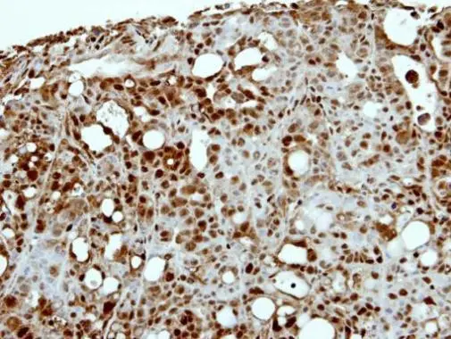
Immunohistochemical analysis of paraffin-embedded NCIN87 xenograft, using CAPG(GTX114301) antibody at 1:500 dilution.
Antigen Retrieval: Trilogy? (EDTA based, pH 8.0) buffer, 15min
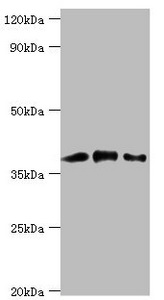
 A: NIH-3T3 10% SDS PAGE GTX114301 diluted at 1:2000")
![CAPG antibody [N3C3] detects CAPG protein at cytoplasm and nucleus by immunofluorescent analysis. Sample: HeLa cells were fixed in 4% paraformaldehyde at RT for 15 min. Green: CAPG protein stained by CAPG antibody [N3C3] (GTX114301) diluted at 1:400. Blue: Hoechst 33342 staining. Scale bar = 10 μm.](https://www.genetex.com/upload/website/prouct_img/normal/GTX114301/GTX114301_40177_20160602_IFA_w_23060501_592.webp "CAPG antibody [N3C3] detects CAPG protein at cytoplasm and nucleus by immunofluorescent analysis. Sample: HeLa cells were fixed in 4% paraformaldehyde at RT for 15 min. Green: CAPG protein stained by CAPG antibody [N3C3] (GTX114301) diluted at 1:400. Blue: Hoechst 33342 staining. Scale bar = 10 μm.")
![Wild-type (WT) and CAPG knockout (KO) HeLa cell extracts (30 μg) were separated by 10% SDS-PAGE, and the membrane was blotted with CAPG antibody [N3C3] (GTX114301) diluted at 1:3000. The HRP-conjugated anti-rabbit IgG antibody (GTX213110-01) was used to detect the primary antibody.](https://www.genetex.com/upload/website/prouct_img/normal/GTX114301/GTX114301_40177_20180810_WB_KO_watermark_w_23060501_571.webp "Wild-type (WT) and CAPG knockout (KO) HeLa cell extracts (30 μg) were separated by 10% SDS-PAGE, and the membrane was blotted with CAPG antibody [N3C3] (GTX114301) diluted at 1:3000. The HRP-conjugated anti-rabbit IgG antibody (GTX213110-01) was used to detect the primary antibody.")
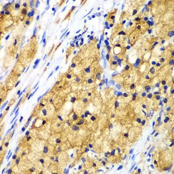
Immunohistochemical analysis of paraffin-embedded NCIN87 xenograft, using CAPG(GTX114301) antibody at 1:500 dilution.
Antigen Retrieval: Trilogy? (EDTA based, pH 8.0) buffer, 15min
CAPG antibody [N3C3]
GTX114301
ApplicationsImmunoFluorescence, Western Blot, ImmunoCytoChemistry, ImmunoHistoChemistry, ImmunoHistoChemistry Paraffin
Product group Antibodies
ReactivityHuman, Mouse
TargetCAPG
Overview
- SupplierGeneTex
- Product NameCAPG antibody [N3C3]
- Delivery Days Customer9
- Application Supplier NoteWB: 1:500-1:3000. ICC/IF: 1:100-1:1000. IHC-P: 1:100-1:1000. *Optimal dilutions/concentrations should be determined by the researcher.Not tested in other applications.
- ApplicationsImmunoFluorescence, Western Blot, ImmunoCytoChemistry, ImmunoHistoChemistry, ImmunoHistoChemistry Paraffin
- CertificationResearch Use Only
- ClonalityPolyclonal
- Concentration1 mg/ml
- ConjugateUnconjugated
- Gene ID822
- Target nameCAPG
- Target descriptioncapping actin protein, gelsolin like
- Target synonymsAFCP, HEL-S-66, MCP, macrophage-capping protein, actin-regulatory protein CAP-G, capping protein (actin filament), gelsolin-like, epididymis secretory protein Li 66, gelsolin-like capping protein
- HostRabbit
- IsotypeIgG
- Protein IDP40121
- Protein NameMacrophage-capping protein
- Scientific DescriptionThis gene encodes a member of the gelsolin/villin family of actin-regulatory proteins. The encoded protein reversibly blocks the barbed ends of F-actin filaments in a Ca2+ and phosphoinositide-regulated manner, but does not sever preformed actin filaments. By capping the barbed ends of actin filaments, the encoded protein contributes to the control of actin-based motility in non-muscle cells. Alternatively spliced transcript variants have been observed, but have not been fully described. [provided by RefSeq]
- ReactivityHuman, Mouse
- Storage Instruction-20°C or -80°C,2°C to 8°C
- UNSPSC41116161
Datasheet
Related products
Product group Antibodies
Anti-CAPG AntibodyA25015
ApplicationsWestern Blot, ImmunoHistoChemistry
ReactivityHuman, Mouse, Rat
- SizePrice
Product group Antibodies
Anti-CAPG Antibody144-07324
ApplicationsWestern Blot, ImmunoHistoChemistry
ReactivityHuman, Mouse, Rat
TargetCAPG
- SizePrice
Product group Antibodies
ApplicationsImmunoFluorescence, ELISA, ImmunoCytoChemistry, ImmunoHistoChemistry, ImmunoHistoChemistry Frozen, ImmunoHistoChemistry Paraffin
ReactivityBovine, Canine, Equine, Human, Mouse, Rat, Sheep
TargetCAPG
- SizePrice
Product group Antibodies
CAPG AntibodyCSB-PA004489ESR2HU
ApplicationsWestern Blot, ELISA
ReactivityHuman, Rat
TargetCAPG
- SizePrice
Product group Antibodies

Goat anti-CAPGEB12421
ApplicationsWestern Blot, ELISA
ReactivityCanine, Human
TargetCAPG
- SizePrice

Product group Antibodies
ApplicationsWestern Blot
ReactivityHuman, Mouse, Rat
TargetCAPG
- SizePrice
Product group Antibodies

CAPG AntibodyLS-C401165
ApplicationsWestern Blot, ELISA, ImmunoHistoChemistry
ReactivityHuman, Mouse, Rat
TargetCAPG
- SizePrice
Product group Antibodies
CAPG antibodyGTX55547
ApplicationsWestern Blot, ImmunoHistoChemistry, ImmunoHistoChemistry Paraffin
ReactivityHuman, Mouse, Rat
TargetCAPG
- SizePrice
Product group Antibodies
TargetCAPG
- SizePrice

Product group Antibodies
Anti-CAPG AntibodyHPA019080
ApplicationsWestern Blot, ImmunoCytoChemistry, ImmunoHistoChemistry
ReactivityHuman
TargetCAPG
- SizePrice