

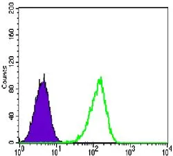
FACS analysis of Lovo cells using GTX80395 CARM1 antibody [3H2]. Green : CARM1 Purple : negative control
![ICC/IF analysis of HeLa cells using GTX80395 CARM1 antibody [3H2]. Green : CARM1 Red: Actin filaments](https://www.genetex.com/upload/website/prouct_img/normal/GTX80395/GTX80395_20170912_ICCIF_w_23061322_186.webp "ICC/IF analysis of HeLa cells using GTX80395 CARM1 antibody [3H2]. Green : CARM1 Red: Actin filaments")
![IHC-P analysis of breast cancer tissue (left) and ovarian cancer tissue (right) using GTX80395 CARM1 antibody [3H2].](https://www.genetex.com/upload/website/prouct_img/normal/GTX80395/GTX80395_20170912_IHC-P_w_23061322_719.webp "IHC-P analysis of breast cancer tissue (left) and ovarian cancer tissue (right) using GTX80395 CARM1 antibody [3H2].")
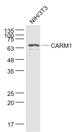
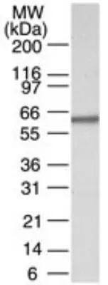
![WB analysis of MCF-7 (1), HeLa (2), NIH3T3 (3), HL-60 (4), LNcap (5), Jurkat (6), PC-3 (7), Cos7 (8), and PC-12 (9) cell lysate using GTX80395 CARM1 antibody [3H2].](https://www.genetex.com/upload/website/prouct_img/normal/GTX80395/GTX80395_20170912_WB_w_23061322_837.webp "WB analysis of MCF-7 (1), HeLa (2), NIH3T3 (3), HL-60 (4), LNcap (5), Jurkat (6), PC-3 (7), Cos7 (8), and PC-12 (9) cell lysate using GTX80395 CARM1 antibody [3H2].")
FACS analysis of Lovo cells using GTX80395 CARM1 antibody [3H2]. Green : CARM1 Purple : negative control
CARM1 antibody [3H2]
GTX80395
ApplicationsFlow Cytometry, ImmunoFluorescence, Western Blot, ELISA, ImmunoCytoChemistry, ImmunoHistoChemistry, ImmunoHistoChemistry Paraffin
Product group Antibodies
ReactivityHuman, Monkey, Mouse, Rat
TargetCARM1
Overview
- SupplierGeneTex
- Product NameCARM1 antibody [3H2]
- Delivery Days Customer9
- Application Supplier NoteWB: 1/500 - 1/2000. ICC/IF: 1/200 - 1/1000. IHC-P: 1/200 - 1/1000. FACS: 1/200 - 1/400. ELISA: 1/10000. *Optimal dilutions/concentrations should be determined by the researcher.Not tested in other applications.
- ApplicationsFlow Cytometry, ImmunoFluorescence, Western Blot, ELISA, ImmunoCytoChemistry, ImmunoHistoChemistry, ImmunoHistoChemistry Paraffin
- CertificationResearch Use Only
- ClonalityMonoclonal
- Clone ID3H2
- ConjugateUnconjugated
- Gene ID10498
- Target nameCARM1
- Target descriptioncoactivator associated arginine methyltransferase 1
- Target synonymsPRMT4, histone-arginine methyltransferase CARM1, protein arginine N-methyltransferase 4
- HostMouse
- IsotypeIgG1
- Protein IDQ86X55
- Protein NameHistone-arginine methyltransferase CARM1
- Scientific DescriptionProtein arginine N-methyltransferases, such as CARM1, catalyze the transfer of a methyl group from S-adenosyl-L-methionine to the side chain nitrogens of arginine residues within proteins to form methylated arginine derivatives and S-adenosyl-L-homocysteine. Protein arginine methylation has been implicated in signal transduction, metabolism of nascent pre-RNA, and transcriptional activation (Frankel et al., 2002 [PubMed 11724789]).[supplied by OMIM, Mar 2008]
- ReactivityHuman, Monkey, Mouse, Rat
- Storage Instruction-20°C or -80°C,2°C to 8°C
- UNSPSC12352203
Datasheet
Related products
Product group Antibodies
Carm1 Polyclonal AntibodyCAC07657
ApplicationsImmunoFluorescence, Western Blot, ELISA, ImmunoHistoChemistry
ReactivityMouse
TargetCARM1
- SizePrice
Product group Antibodies
CARM1 Polyclonal AntibodyBS-4098R
ApplicationsImmunoFluorescence, Western Blot, ELISA, ImmunoCytoChemistry, ImmunoHistoChemistry, ImmunoHistoChemistry Frozen, ImmunoHistoChemistry Paraffin
ReactivityBovine, Canine, Human, Mouse, Porcine, Rat
TargetCARM1
- SizePrice
Product group Antibodies
Anti-CARM1 Antibody144-02246
ApplicationsWestern Blot
ReactivityHuman, Mouse, Rat
TargetCARM1
- SizePrice
Product group Antibodies
Anti-PRMT4 AntibodyA97327
ApplicationsWestern Blot, ELISA
ReactivityHuman, Mouse, Rat
- SizePrice
Product group Antibodies
CARM1 antibodyGTX78920
ApplicationsWestern Blot
ReactivityHuman
TargetCARM1
- SizePrice
Product group Antibodies
CARM1 (phospho Ser228) antibodyGTX79118
ApplicationsWestern Blot
ReactivityHuman
TargetCARM1
- SizePrice
Product group Antibodies
CARM1 antibodyGTX31871
ApplicationsImmunoFluorescence, Western Blot, ELISA, ImmunoCytoChemistry
ReactivityHuman, Mouse, Rat
TargetCARM1
- SizePrice
Product group Antibodies
CARM1 antibodyGTX129040
ApplicationsWestern Blot, ImmunoHistoChemistry, ImmunoHistoChemistry Paraffin
ReactivityHuman
TargetCARM1
- SizePrice
Product group Antibodies
CARM1 antibodyGTX13707
ApplicationsWestern Blot
ReactivityHuman, Mouse, Rat, Xenopus
TargetCARM1
- SizePrice