

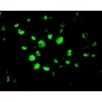
ICC/IF analysis of HeLa cells using GTX22012 Caspase 10 antibody. Green : Primary antibody Blue : DAPI Dilution : 20 μg/ml
, Jurkat (J), A431 (A), K562 (K) whole cell lysates with Caspase-10 antibody (GTX22012) at 1μg/ml.")
ICC/IF analysis of HeLa cells using GTX22012 Caspase 10 antibody. Green : Primary antibody Blue : DAPI Dilution : 20 μg/ml
Caspase 10 antibody
GTX22012
ApplicationsImmunoFluorescence, Western Blot, ELISA, ImmunoCytoChemistry
Product group Antibodies
ReactivityHuman
TargetCASP10
Overview
- SupplierGeneTex
- Product NameCaspase 10 antibody
- Delivery Days Customer9
- Application Supplier NoteWB: 1microg/ml. *Optimal dilutions/concentrations should be determined by the researcher.Not tested in other applications.
- ApplicationsImmunoFluorescence, Western Blot, ELISA, ImmunoCytoChemistry
- CertificationResearch Use Only
- ClonalityPolyclonal
- ConjugateUnconjugated
- Gene ID843
- Target nameCASP10
- Target descriptioncaspase 10
- Target synonymsALPS2, FLICE-2, FLICE2, MCH4, caspase-10, CASP-10, FADD-like ICE2, FAS-associated death domain protein interleukin-1B-converting enzyme 2, ICE-like apoptotic protease 4, apoptotic protease MCH-4, caspase 10 apoptosis-related cysteine peptidase, caspase 10, apoptosis-related cysteine protease, interleukin-1B-converting enzyme 2
- HostRabbit
- IsotypeIgG
- Protein IDQ92851
- Protein NameCaspase-10
- Scientific DescriptionThis gene encodes a protein which is a member of the cysteine-aspartic acid protease (caspase) family. Sequential activation of caspases plays a central role in the execution-phase of cell apoptosis. Caspases exist as inactive proenzymes which undergo proteolytic processing at conserved aspartic residues to produce two subunits, large and small, that dimerize to form the active enzyme. This protein cleaves and activates caspases 3 and 7, and the protein itself is processed by caspase 8. Mutations in this gene are associated with type IIA autoimmune lymphoproliferative syndrome, non-Hodgkin lymphoma and gastric cancer. Alternatively spliced transcript variants encoding different isoforms have been described for this gene. [provided by RefSeq, Apr 2011]
- ReactivityHuman
- Storage Instruction-20°C or -80°C,2°C to 8°C
- UNSPSC41116161
Datasheet
Related products

Product group Antibodies
Anti-Caspase 10 AntibodyA96513
ApplicationsImmunoFluorescence, Western Blot, ELISA, ImmunoHistoChemistry
ReactivityHuman
- SizePrice
Product group Antibodies
Anti-CASP10 Antibody144-60015
ApplicationsImmunoFluorescence, Western Blot
ReactivityHuman
TargetCASP10
- SizePrice

Product group Antibodies
Anti-Caspase-10/CASP10 Antibody Picoband(r)A02190-2-CARRIER-FREE
ApplicationsFlow Cytometry, Western Blot, ELISA
ReactivityHuman
TargetCASP10
- SizePrice
Product group Antibodies
Caspase-10 Recombinant Antibody, Biotin ConjugatedBSM-61328R-BIOTIN
ApplicationsImmunoPrecipitation, Western Blot, ImmunoHistoChemistry, ImmunoHistoChemistry Frozen, ImmunoHistoChemistry Paraffin
TargetCASP10
- SizePrice

Product group Antibodies
CASP10 AntibodyCSB-PA001228
ApplicationsImmunoFluorescence, Western Blot, ELISA, ImmunoHistoChemistry
ReactivityHuman
- SizePrice
Product group Antibodies
ApplicationsImmunoPrecipitation, Western Blot, ImmunoCytoChemistry, ImmunoHistoChemistry
ReactivityMouse, Porcine
TargetCASP10
- SizePrice
Product group Antibodies

CASP10 / Caspase 10 AntibodyLS-C403736
ApplicationsELISA, ImmunoHistoChemistry
ReactivityHuman
TargetCASP10
- SizePrice
Product group Antibodies
Caspase 10 antibodyGTX10452
ApplicationsWestern Blot, ImmunoHistoChemistry
ReactivityHuman
TargetCASP10
- SizePrice
Product group Antibodies
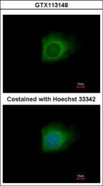
Caspase 10 antibodyGTX113148
ApplicationsImmunoFluorescence, Western Blot, ImmunoCytoChemistry, ImmunoHistoChemistry, ImmunoHistoChemistry Paraffin
ReactivityHuman
TargetCASP10
- SizePrice