

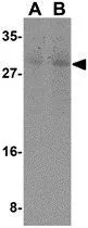
WB analysis of mouse skeletal muscle tissue lysate using GTX31705 Caspase 7 antibody. Working concentration : (A) 0.5 and (B) 1 μg/ml

WB analysis of mouse skeletal muscle tissue lysate using GTX31705 Caspase 7 antibody. Working concentration : (A) 0.5 and (B) 1 μg/ml
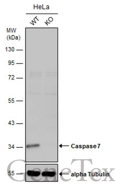
Caspase 7 antibody
GTX31705
ApplicationsWestern Blot, ELISA, ImmunoHistoChemistry, ImmunoHistoChemistry Paraffin
Product group Antibodies
ReactivityHuman, Mouse, Rat
TargetCASP7
Overview
- SupplierGeneTex
- Product NameCaspase 7 antibody
- Delivery Days Customer9
- Application Supplier NoteWB: 0.5 - 1 microg/mL. IHC-P: 10 microg/mL. *Optimal dilutions/concentrations should be determined by the researcher.Not tested in other applications.
- ApplicationsWestern Blot, ELISA, ImmunoHistoChemistry, ImmunoHistoChemistry Paraffin
- CertificationResearch Use Only
- ClonalityPolyclonal
- Concentration1 mg/ml
- ConjugateUnconjugated
- Gene ID840
- Target nameCASP7
- Target descriptioncaspase 7
- Target synonymsCASP-7, CMH-1, ICE-LAP3, LICE2, MCH3, caspase-7, ICE-like apoptotic protease 3, apoptotic protease MCH-3, caspase 7, apoptosis-related cysteine peptidase, caspase 7, apoptosis-related cysteine protease
- HostRabbit
- IsotypeIgG
- Protein IDP55210
- Protein NameCaspase-7
- Scientific DescriptionThis gene encodes a member of the cysteine-aspartic acid protease (caspase) family. Sequential activation of caspases plays a central role in the execution-phase of cell apoptosis. Caspases exist as inactive proenzymes which undergo proteolytic processing at conserved aspartic residues to produce two subunits, large and small, that dimerize to form the active enzyme. The precursor of the encoded protein is cleaved by caspase 3 and 10, is activated upon cell death stimuli and induces apoptosis. Alternatively spliced transcript variants encoding multiple isoforms have been observed for this gene. [provided by RefSeq, May 2012]
- ReactivityHuman, Mouse, Rat
- Storage Instruction-20°C or -80°C,2°C to 8°C
- UNSPSC41116161
Datasheet
Related products

Product group Antibodies
Anti-Caspase-7 AntibodyA98572
ApplicationsWestern Blot, ELISA, ImmunoHistoChemistry
ReactivityHuman
- SizePrice

Product group Antibodies
Anti-Caspase-7/CASP7 Antibody Picoband(r)A01044-2-CARRIER-FREE
ApplicationsFlow Cytometry, ImmunoFluorescence, Western Blot, ELISA, ImmunoCytoChemistry, ImmunoHistoChemistry
ReactivityHuman, Rat
TargetCASP7
- SizePrice
Product group Antibodies

CASP7 / Caspase 7 AntibodyLS-C746750
ApplicationsImmunoPrecipitation, Western Blot, ImmunoHistoChemistry
ReactivityHuman, Mouse, Rat
TargetCASP7
- SizePrice
Product group Antibodies
Caspase-7 Recombinant AntibodyBSM-60304R
ApplicationsImmunoFluorescence, Western Blot, ImmunoHistoChemistry, ImmunoHistoChemistry Frozen, ImmunoHistoChemistry Paraffin
ReactivityHuman, Mouse
TargetCASP7
- SizePrice
Product group Antibodies
CASP7 AntibodyCSB-PA001233
ApplicationsWestern Blot, ELISA, ImmunoHistoChemistry
ReactivityHuman
TargetCASP7
- SizePrice
Product group Antibodies
CASP7 Polyclonal AntibodyCAC14141
ApplicationsImmunoFluorescence, ImmunoPrecipitation, Western Blot, ELISA, ImmunoHistoChemistry
TargetCASP7
- SizePrice
![WB analysis of HeLa cell lysate using GTX30242 Caspase 7 antibody[Mch3 1-1-11].](https://www.genetex.com/upload/website/prouct_img/normal/GTX30242/GTX30242_1373_WB_w_23060722_903.webp)
Product group Antibodies
Caspase 7 antibody [Mch3 1-1-11]GTX30242
ApplicationsWestern Blot
ReactivityHuman, Mouse, Rat
TargetCASP7
- SizePrice
Product group Antibodies
References
Caspase 7 antibodyGTX31704
ApplicationsWestern Blot, ELISA, ImmunoHistoChemistry, ImmunoHistoChemistry Paraffin
ReactivityHuman, Mouse, Rat
TargetCASP7
- SizePrice
Product group Antibodies
References
ApplicationsWestern Blot, ImmunoHistoChemistry, ImmunoHistoChemistry Paraffin
ReactivityHuman
TargetCASP7
- SizePrice
Product group Antibodies
Caspase 7 antibodyGTX123679
ApplicationsImmunoPrecipitation, Western Blot, ImmunoHistoChemistry, ImmunoHistoChemistry Paraffin
ReactivityHuman, Rat
TargetCASP7
- SizePrice