

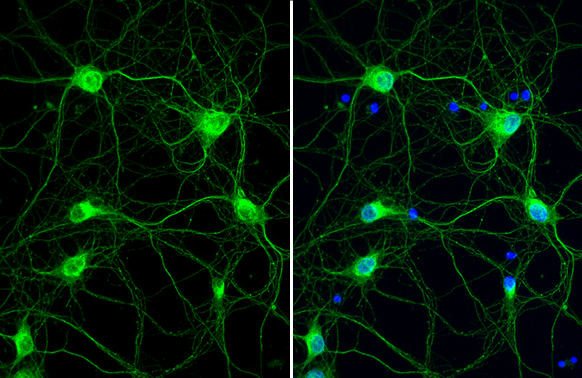
Cathepsin S antibody [HL2302] detects Cathepsin S protein at fiber by immunofluorescent analysis. Sample: DIV9 rat E18 primary cortical neuron cells were fixed in 4% paraformaldehyde at RT for 15 min. Green: Cathepsin S stained by Cathepsin S antibody [HL2302] (GTX638369) diluted at 1:250. Blue: Fluoroshield with DAPI (GTX30920).
![Cathepsin S antibody [HL2302] detects Cathepsin S protein at cytoplasmic vesicle by immunofluorescent analysis. Sample: U87-MG cells were fixed in ice-cold MeOH for 5 min. Green: Cathepsin S stained by Cathepsin S antibody [HL2302] (GTX638369) diluted at 1:500. Blue: Fluoroshield with DAPI (GTX30920).](https://www.genetex.com/upload/website/prouct_img/normal/GTX638369/GTX638369_T-44991_20230512_ICC_IF_23060622_487.webp "Cathepsin S antibody [HL2302] detects Cathepsin S protein at cytoplasmic vesicle by immunofluorescent analysis. Sample: U87-MG cells were fixed in ice-cold MeOH for 5 min. Green: Cathepsin S stained by Cathepsin S antibody [HL2302] (GTX638369) diluted at 1:500. Blue: Fluoroshield with DAPI (GTX30920).")
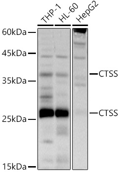
![Various whole cell extracts (30 μg) were separated by 12% SDS-PAGE, and the membrane was blotted with Cathepsin S antibody [HL2302] (GTX638369) diluted at 1:1000. The HRP-conjugated anti-rabbit IgG antibody (GTX213110-01) was used to detect the primary antibody. Corresponding RNA expression data for the same cell lines are based on Human Protein Atlas program.](https://www.genetex.com/upload/website/prouct_img/normal/GTX638369/GTX638369_45110_20230721_WB_TPM_watermark_23072519_907.webp "Various whole cell extracts (30 μg) were separated by 12% SDS-PAGE, and the membrane was blotted with Cathepsin S antibody [HL2302] (GTX638369) diluted at 1:1000. The HRP-conjugated anti-rabbit IgG antibody (GTX213110-01) was used to detect the primary antibody. Corresponding RNA expression data for the same cell lines are based on Human Protein Atlas program.")
![Cathepsin S antibody [HL2302] detects Cathepsin S protein by immunohistochemical analysis. Sample: Paraffin-embedded mouse tissues. Cathepsin S stained by Cathepsin S antibody [HL2302] (GTX638369) diluted at 1:100. Antigen Retrieval: Citrate buffer, pH 6.0, 15 min](https://www.genetex.com/upload/website/prouct_img/normal/GTX638369/GTX638369_T-44991_20230801_IHC-P_multiple_M_23081619_160.webp "Cathepsin S antibody [HL2302] detects Cathepsin S protein by immunohistochemical analysis. Sample: Paraffin-embedded mouse tissues. Cathepsin S stained by Cathepsin S antibody [HL2302] (GTX638369) diluted at 1:100. Antigen Retrieval: Citrate buffer, pH 6.0, 15 min")
![Cathepsin S antibody [HL2302] detects Cathepsin S protein at cytoplasm by immunohistochemical analysis. Sample: Paraffin-embedded human tissue. Cathepsin S stained by Cathepsin S antibody [HL2302] (GTX638369) diluted at 1:100. Antigen Retrieval: Citrate buffer, pH 6.0, 15 min](https://www.genetex.com/upload/website/prouct_img/normal/GTX638369/GTX638369_45110_20230928_IHC-P_multiple_23100319_546.webp "Cathepsin S antibody [HL2302] detects Cathepsin S protein at cytoplasm by immunohistochemical analysis. Sample: Paraffin-embedded human tissue. Cathepsin S stained by Cathepsin S antibody [HL2302] (GTX638369) diluted at 1:100. Antigen Retrieval: Citrate buffer, pH 6.0, 15 min")
Cathepsin S antibody [HL2302] detects Cathepsin S protein at fiber by immunofluorescent analysis. Sample: DIV9 rat E18 primary cortical neuron cells were fixed in 4% paraformaldehyde at RT for 15 min. Green: Cathepsin S stained by Cathepsin S antibody [HL2302] (GTX638369) diluted at 1:250. Blue: Fluoroshield with DAPI (GTX30920).
Cathepsin S antibody [HL2302]
GTX638369
ApplicationsImmunoFluorescence, Western Blot, ImmunoCytoChemistry, ImmunoHistoChemistry, ImmunoHistoChemistry Paraffin
Product group Antibodies
ReactivityHuman, Mouse, Rat
TargetCTSS
Overview
- SupplierGeneTex
- Product NameCathepsin S antibody [HL2302]
- Delivery Days Customer9
- Application Supplier NoteWB: 1:500-1:3000. *Optimal dilutions/concentrations should be determined by the researcher.Not tested in other applications.
- ApplicationsImmunoFluorescence, Western Blot, ImmunoCytoChemistry, ImmunoHistoChemistry, ImmunoHistoChemistry Paraffin
- CertificationResearch Use Only
- ClonalityMonoclonal
- Clone IDHL2302
- Concentration1 mg/ml
- ConjugateUnconjugated
- Gene ID1520
- Target nameCTSS
- Target descriptioncathepsin S
- Target synonymscathepsin S
- HostRabbit
- IsotypeIgG
- Protein IDP25774
- Protein NameCathepsin S
- Scientific DescriptionThe protein encoded by this gene, a member of the peptidase C1 family, is a lysosomal cysteine proteinase that may participate in the degradation of antigenic proteins to peptides for presentation on MHC class II molecules. The encoded protein can function as an elastase over a broad pH range in alveolar macrophages. Alternatively spliced transcript variants encoding distinct isoforms have been found for this gene. [provided by RefSeq, Dec 2010]
- ReactivityHuman, Mouse, Rat
- Storage Instruction-20°C or -80°C,2°C to 8°C
- UNSPSC41116161
Datasheet
Related products
Product group Antibodies
ApplicationsImmunoFluorescence, Western Blot, ImmunoCytoChemistry
ReactivityHuman, Mouse, Rat
- SizePrice
Product group Antibodies

Anti-Cathepsin S [Fsn0503 (1E4)]Ab02999-1.1
ApplicationsImmunoFluorescence, Western Blot, ELISA, ImmunoCytoChemistry, ImmunoHistoChemistry, Neutralisation/Blocking, Other Application
ReactivityHuman
TargetCTSS
- SizePrice

Product group Antibodies
Anti-CTSS AntibodyHPA002988
ApplicationsWestern Blot, ImmunoCytoChemistry, ImmunoHistoChemistry
ReactivityHuman
TargetCTSS
- SizePrice
Product group Antibodies
CTSS AntibodyCSB-PA10729A0RB
ApplicationsWestern Blot, ELISA, ImmunoHistoChemistry
ReactivityHuman
TargetCTSS
- SizePrice
Product group Antibodies

CTSS / Cathepsin S AntibodyLS-C484038
ApplicationsWestern Blot, ELISA
ReactivityMouse
TargetCTSS
- SizePrice
Product group Antibodies
Cathepsin S Recombinant Antibody, AbBy Fluor-594 ConjugatedBSM-61770R-BF594
ApplicationsImmunoFluorescence, Western Blot
ReactivityHuman
TargetCTSS
- SizePrice
Product group Antibodies
Ctss Polyclonal AntibodyCAC09131
ApplicationsWestern Blot, ELISA, ImmunoHistoChemistry
TargetCTSS
- SizePrice
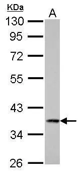
![Whole cell extract (30 μg) was separated by 15% SDS-PAGE, and the membrane was blotted with Cathepsin S antibody [HL2462] (GTX638780) diluted at 1:1000. The HRP-conjugated anti-rabbit IgG antibody (GTX213110-01) was used to detect the primary antibody.](https://www.genetex.com/upload/website/prouct_img/normal/GTX638780/GTX638780_T-45089_20230714_WB_23071822_292.webp)
Product group Antibodies
Cathepsin S antibody [HL2462]GTX638780
ApplicationsWestern Blot, ImmunoHistoChemistry, ImmunoHistoChemistry Paraffin
ReactivityHuman
TargetCTSS
- SizePrice
Product group Antibodies
Cathepsin S antibodyGTX103988
ApplicationsWestern Blot, ImmunoHistoChemistry, ImmunoHistoChemistry Paraffin
ReactivityHuman, Rat
TargetCTSS
- SizePrice