

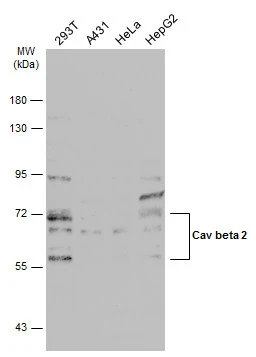
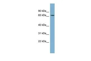
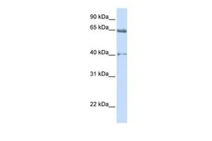
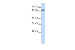
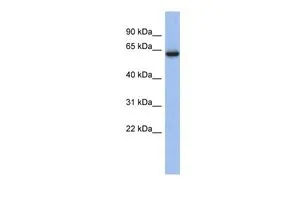
Various whole cell extracts (30 μg) was separated by 7.5% SDS-PAGE, and the membrane was blotted with Cav beta 2 antibody (GTX131906) diluted at 1:1000. The HRP-conjugated anti-rabbit IgG antibody (GTX213110-01) was used to detect the primary antibody.
![Cav beta 2 antibody detects Cav beta 2 protein by immunohistochemical analysis. Sample: Frozen-sectioned mouse mouse cerebellum. Green: Cav beta 2 stained by Cav beta 2 antibody (GTX131906) diluted at 1:250. Red: NF-H, stained by NF-H antibody [GT114] (GTX634289) diluted at 1:500. Blue: Fluoroshield with DAPI (GTX30920). Antigen Retrieval: Citrate buffer, pH 6.0, 10 min](https://www.genetex.com/upload/website/prouct_img/normal/GTX131906/GTX131906_43103_20180530_IHC-Fr_M_2_w_23051500_429.webp "Cav beta 2 antibody detects Cav beta 2 protein by immunohistochemical analysis. Sample: Frozen-sectioned mouse mouse cerebellum. Green: Cav beta 2 stained by Cav beta 2 antibody (GTX131906) diluted at 1:250. Red: NF-H, stained by NF-H antibody [GT114] (GTX634289) diluted at 1:500. Blue: Fluoroshield with DAPI (GTX30920). Antigen Retrieval: Citrate buffer, pH 6.0, 10 min")
Various whole cell extracts (30 μg) was separated by 7.5% SDS-PAGE, and the membrane was blotted with Cav beta 2 antibody (GTX131906) diluted at 1:1000. The HRP-conjugated anti-rabbit IgG antibody (GTX213110-01) was used to detect the primary antibody.
Cav beta 2 antibody
GTX131906
ApplicationsWestern Blot, ImmunoHistoChemistry, ImmunoHistoChemistry Frozen
Product group Antibodies
ReactivityHuman, Mouse
TargetCACNB2
Overview
- SupplierGeneTex
- Product NameCav beta 2 antibody
- Delivery Days Customer9
- Application Supplier NoteWB: 1:500-1:3000. IHC-Fr: 1:100-1:1000. *Optimal dilutions/concentrations should be determined by the researcher.Not tested in other applications.
- ApplicationsWestern Blot, ImmunoHistoChemistry, ImmunoHistoChemistry Frozen
- CertificationResearch Use Only
- ClonalityPolyclonal
- Concentration1.66 mg/ml
- ConjugateUnconjugated
- Gene ID783
- Target nameCACNB2
- Target descriptioncalcium voltage-gated channel auxiliary subunit beta 2
- Target synonymsCAB2, CACNLB2, CAVB2, MYSB, voltage-dependent L-type calcium channel subunit beta-2, calcium channel voltage-dependent subunit beta 2, calcium channel, voltage-dependent, beta 2 subunit, lambert-Eaton myasthenic syndrome antigen B, myasthenic (Lambert-Eaton) syndrome antigen B
- HostRabbit
- IsotypeIgG
- Protein IDQ08289
- Protein NameVoltage-dependent L-type calcium channel subunit beta-2
- Scientific DescriptionThis gene encodes a subunit of a voltage-dependent calcium channel protein that is a member of the voltage-gated calcium channel superfamily. The gene product was originally identified as an antigen target in Lambert-Eaton myasthenic syndrome, an autoimmune disorder. Mutations in this gene are associated with Brugada syndrome. Alternatively spliced variants encoding different isoforms have been described. [provided by RefSeq, Feb 2013]
- ReactivityHuman, Mouse
- Storage Instruction-20°C or -80°C,2°C to 8°C
- UNSPSC41116161
Datasheet
Related products
Product group Antibodies
CACNB2 AntibodyCSB-PA191134
ApplicationsELISA, ImmunoHistoChemistry
ReactivityHuman, Mouse, Rat
TargetCACNB2
- SizePrice
Product group Antibodies
Anti-CACNB2 AntibodyA91433
ApplicationsWestern Blot, ImmunoHistoChemistry
ReactivityHuman, Mouse, Rat
- SizePrice
Product group Antibodies

Goat anti-CACNB2 (aa565-579)EB11178
ApplicationsWestern Blot, ELISA
ReactivityBovine, Canine, Human, Mouse, Rat
TargetCACNB2
- SizePrice

Product group Antibodies
Anti-CACNB2 AntibodyHPA035326
ApplicationsImmunoHistoChemistry
ReactivityHuman
TargetCACNB2
- SizePrice
Product group Antibodies

CACNB2 AntibodyLS-C406259
ApplicationsELISA, ImmunoHistoChemistry
ReactivityHuman, Mouse, Rat
TargetCACNB2
- SizePrice
Product group Antibodies
Cav beta 2 antibody, InternalGTX47534
ApplicationsWestern Blot
ReactivityHuman
TargetCACNB2
- SizePrice
Product group Antibodies
Cav beta 2 antibody, InternalGTX47535
ApplicationsWestern Blot
ReactivityHuman
TargetCACNB2
- SizePrice
Product group Antibodies
Cav beta 2 antibody, InternalGTX47537
ApplicationsWestern Blot
ReactivityHuman
TargetCACNB2
- SizePrice
Product group Antibodies
Cav beta 2 antibody, InternalGTX47538
ApplicationsWestern Blot
ReactivityHuman
TargetCACNB2
- SizePrice
Product group Antibodies
Cav beta 2 antibody, InternalGTX47609
ApplicationsWestern Blot
ReactivityHuman
TargetCACNB2
- SizePrice