

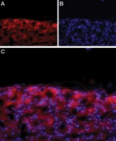
IHC-Fr analysis of rat DRG tissue using GTX16633 Cav1.3 antibody. Panel A : CaV1.3 labeling (red) appears in the cell bodies of the DRG. Panel B : Nuclear staining using DAPI as the counterstain. Panel C : Merged image of A and B.

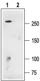
, mouse brain (lanes 2 and 5) and C6 (lanes 3 and 6) lysates using GTX16633 Cav1.3 antibody preincubated with or without immunogen peptide. Dilution : 1:200")
IHC-Fr analysis of rat DRG tissue using GTX16633 Cav1.3 antibody. Panel A : CaV1.3 labeling (red) appears in the cell bodies of the DRG. Panel B : Nuclear staining using DAPI as the counterstain. Panel C : Merged image of A and B.
Cav1.3 antibody
GTX16633
ApplicationsImmunoFluorescence, Western Blot, ImmunoCytoChemistry, ImmunoHistoChemistry, ImmunoHistoChemistry Frozen, Other Application
Product group Antibodies
ReactivityHuman, Mouse, Rat
TargetCacna1d
Overview
- SupplierGeneTex
- Product NameCav1.3 antibody
- Delivery Days Customer7
- ApplicationsImmunoFluorescence, Western Blot, ImmunoCytoChemistry, ImmunoHistoChemistry, ImmunoHistoChemistry Frozen, Other Application
- CertificationResearch Use Only
- ClonalityPolyclonal
- Concentration0.9 mg/ml
- ConjugateUnconjugated
- Gene ID29716
- Target nameCacna1d
- Target descriptioncalcium voltage-gated channel subunit alpha1 D
- Target synonymsvoltage-dependent L-type calcium channel subunit alpha-1D, CaV1.3alpha1, RBD, brain class D, calcium channel alpha-1 subunit, calcium channel, L type, alpha-1 polypeptide, calcium channel, voltage-dependent, L type, alpha 1D subunit, voltage-dependent calcium channel subunit alpha1D, voltage-gated calcium channel pore forming subunit CaV1.3alpha1 IVS3-IVS4 extracellular linker, voltage-gated calcium channel subunit alpha Cav1.3
- HostRabbit
- IsotypeIgG
- Protein IDP27732
- Protein NameVoltage-dependent L-type calcium channel subunit alpha-1D
- Scientific Descriptionhas high voltage L-type calcium channel activity that is sensitive to dihydropyridines [RGD, Feb 2006]
- ReactivityHuman, Mouse, Rat
- Storage Instruction-20°C or -80°C,2°C to 8°C
- UNSPSC41116161
Datasheet
Related products
Product group Antibodies

Cav1.3 AntibodyABX444956
ApplicationsImmunoFluorescence, ImmunoPrecipitation, Western Blot, ImmunoCytoChemistry, ImmunoHistoChemistry, Other Application
- SizePrice
Product group Antibodies
References
Cav1.3 antibodyGTX54755
ApplicationsImmunoFluorescence, ImmunoPrecipitation, Western Blot, ImmunoCytoChemistry, ImmunoHistoChemistry, ImmunoHistoChemistry Frozen
ReactivityHuman, Mouse, Rat
TargetCacna1d
- SizePrice
Product group Antibodies
Cav1.3 antibody [S48A-9]GTX41982
ApplicationsImmunoFluorescence, ImmunoPrecipitation, Western Blot, ImmunoCytoChemistry
ReactivityHuman, Mouse, Rat
TargetCacna1d
- SizePrice