

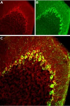
IHC-Fr analysis of mouse cerebellum tissue using GTX54813 Cav3.2 antibody. Panel A : CaV3.2 appears adjacent to Purkinje cells and in fibers in the molecular layer (red). Panel B : Staining of Purkinje cells with mouse anti-parvalbumin (PV, green). Panel C : Merged image of panels A and B demonstrates presence of CaV3.2 adjacent to Purkinje? cells. Dilution : 1:100


IHC-Fr analysis of mouse cerebellum tissue using GTX54813 Cav3.2 antibody. Panel A : CaV3.2 appears adjacent to Purkinje cells and in fibers in the molecular layer (red). Panel B : Staining of Purkinje cells with mouse anti-parvalbumin (PV, green). Panel C : Merged image of panels A and B demonstrates presence of CaV3.2 adjacent to Purkinje? cells. Dilution : 1:100
Cav3.2 antibody
GTX54813
ApplicationsImmunoFluorescence, ImmunoPrecipitation, Western Blot, ImmunoCytoChemistry, ImmunoHistoChemistry, ImmunoHistoChemistry Frozen
Product group Antibodies
ReactivityHuman, Mouse, Rat
TargetCacna1h
Overview
- SupplierGeneTex
- Product NameCav3.2 antibody
- Delivery Days Customer7
- ApplicationsImmunoFluorescence, ImmunoPrecipitation, Western Blot, ImmunoCytoChemistry, ImmunoHistoChemistry, ImmunoHistoChemistry Frozen
- CertificationResearch Use Only
- ClonalityPolyclonal
- Concentration0.8 mg/ml
- ConjugateUnconjugated
- Gene ID114862
- Target nameCacna1h
- Target descriptioncalcium voltage-gated channel subunit alpha1 H
- Target synonymsvoltage-dependent T-type calcium channel subunit alpha-1H, calcium channel, voltage-dependent, T type, alpha 1H subunit, voltage-gated calcium channel subunit alpha Cav3.2
- HostRabbit
- IsotypeIgG
- Protein IDQ9EQ60
- Protein NameVoltage-dependent T-type calcium channel subunit alpha-1H
- Scientific Descriptionacts as a low voltage gated calcium channel; conducts calcium current; involved in neuronal physiology [RGD, Feb 2006]
- ReactivityHuman, Mouse, Rat
- Storage Instruction-20°C or -80°C,2°C to 8°C
- UNSPSC41116161