

CD10 antibody [N2C1], Internal detects MME protein by western blot analysis. A. 50 μg rat kidney lysate/extract 7.5% SDS-PAGE CD10 antibody [N2C1], Internal (GTX111680) dilution: 1:500 The HRP-conjugated anti-rabbit IgG antibody (GTX213110-01) was used to detect the primary antibody.
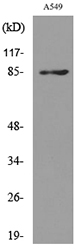
![Various whole cell extracts (30 μg) were separated by 7.5% SDS-PAGE, and the membrane was blotted with CD10 antibody [N2C1], Internal (GTX111680) diluted at 1:1000. The HRP-conjugated anti-rabbit IgG antibody (GTX213110-01) was used to detect the primary antibody.](https://www.genetex.com/upload/website/prouct_img/normal/GTX111680/GTX111680_43411_20181207_WB_w_23060500_335.webp "Various whole cell extracts (30 μg) were separated by 7.5% SDS-PAGE, and the membrane was blotted with CD10 antibody [N2C1], Internal (GTX111680) diluted at 1:1000. The HRP-conjugated anti-rabbit IgG antibody (GTX213110-01) was used to detect the primary antibody.")
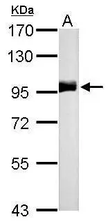
![Various tissue extracts (50 μg) were separated by 7.5% SDS-PAGE, and the membrane was blotted with CD10 antibody [N2C1], Internal (GTX111680) diluted at 1:500. The HRP-conjugated anti-rabbit IgG antibody (GTX213110-01) was used to detect the primary antibody.](https://www.genetex.com/upload/website/prouct_img/normal/GTX111680/GTX111680_43411_20210507_WB_M_R_w_23060500_967.webp "Various tissue extracts (50 μg) were separated by 7.5% SDS-PAGE, and the membrane was blotted with CD10 antibody [N2C1], Internal (GTX111680) diluted at 1:500. The HRP-conjugated anti-rabbit IgG antibody (GTX213110-01) was used to detect the primary antibody.")
CD10 antibody [N2C1], Internal detects MME protein by western blot analysis. A. 50 μg rat kidney lysate/extract 7.5% SDS-PAGE CD10 antibody [N2C1], Internal (GTX111680) dilution: 1:500 The HRP-conjugated anti-rabbit IgG antibody (GTX213110-01) was used to detect the primary antibody.
CD10 antibody [N2C1], Internal
GTX111680
ApplicationsWestern Blot
Product group Antibodies
ReactivityHuman, Mouse, Rat
TargetMME
Overview
- SupplierGeneTex
- Product NameCD10 antibody [N2C1], Internal
- Delivery Days Customer9
- Application Supplier NoteWB: 1:500-1:3000. *Optimal dilutions/concentrations should be determined by the researcher.Not tested in other applications.
- ApplicationsWestern Blot
- CertificationResearch Use Only
- ClonalityPolyclonal
- Concentration1.01 mg/ml
- ConjugateUnconjugated
- Gene ID4311
- Target nameMME
- Target descriptionmembrane metalloendopeptidase
- Target synonymsCALLA, CD10, CMT2T, NEP, SCA43, SFE, neprilysin, atriopeptidase, common acute lymphocytic leukemia antigen, membrane metallo-endopeptidase (neutral endopeptidase, enkephalinase, CALLA, CD10), neprilysin-390, neprilysin-411, neutral endopeptidase 24.11, skin fibroblast elastase
- HostRabbit
- IsotypeIgG
- Protein IDP08473
- Protein NameNeprilysin
- Scientific DescriptionThis gene encodes a common acute lymphocytic leukemia antigen that is an important cell surface marker in the diagnosis of human acute lymphocytic leukemia (ALL). This protein is present on leukemic cells of pre-B phenotype, which represent 85% of cases of ALL. This protein is not restricted to leukemic cells, however, and is found on a variety of normal tissues. It is a glycoprotein that is particularly abundant in kidney, where it is present on the brush border of proximal tubules and on glomerular epithelium. The protein is a neutral endopeptidase that cleaves peptides at the amino side of hydrophobic residues and inactivates several peptide hormones including glucagon, enkephalins, substance P, neurotensin, oxytocin, and bradykinin. This gene, which encodes a 100-kD type II transmembrane glycoprotein, exists in a single copy of greater than 45 kb. The 5 untranslated region of this gene is alternatively spliced, resulting in four separate mRNA transcripts. The coding region is not affected by alternative splicing. [provided by RefSeq]
- ReactivityHuman, Mouse, Rat
- Storage Instruction-20°C or -80°C,2°C to 8°C
- UNSPSC41116161
Datasheet
Related products
Product group Antibodies
Anti-MME AntibodyA98473
ApplicationsWestern Blot, ELISA
ReactivityHuman, Mouse, Rat
- SizePrice

Product group Antibodies
Anti-MME AntibodyAMAB91788
ApplicationsImmunoHistoChemistry
ReactivityHuman
TargetMME
- SizePrice
Product group Antibodies

MME / CD10 Antibody (clone CB-CALLA)LS-C769989
ApplicationsFlow Cytometry
ReactivityHuman
TargetMME
- SizePrice
Product group Antibodies

Anti-CD10 [NL-1]Ab00950-10.0
ApplicationsImmunoFluorescence, ImmunoPrecipitation, Western Blot
ReactivityHuman
TargetMME
- SizePrice
Product group Antibodies
References
CD10 Polyclonal AntibodyBS-0527R
ApplicationsWestern Blot
ReactivityHuman, Mouse, Rat
TargetMME
- SizePrice
Product group Antibodies
MME Monoclonal AntibodyCSB-MA000230
ApplicationsELISA, ImmunoHistoChemistry
ReactivityHuman, Mouse, Rat
TargetMME
- SizePrice
Product group Antibodies
ApplicationsFlow Cytometry
TargetMME
- SizePrice
![IHC-P analysis of human renal cell carcinoma tissue using GTX17135 CD10 antibody [56C6] (ready-to-use).](https://www.genetex.com/upload/website/prouct_img/normal/GTX17135/GTX17135_20191203_IHC-P_27_w_23060620_921.webp)
Product group Antibodies
ApplicationsImmunoHistoChemistry, ImmunoHistoChemistry Paraffin
ReactivityHuman
TargetMME
- SizePrice