

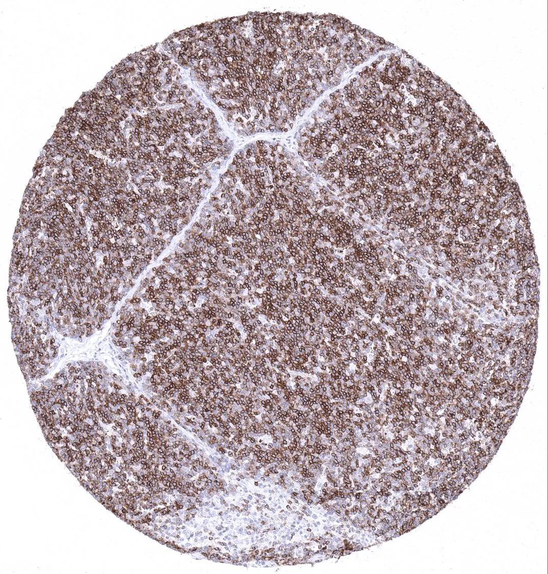
IHC-P analysis of human thymic cortex tissue using GTX04407 CD1a antibody [MSVA-001M] HistoMAX?. A strong CD1a immunostaining is seen in 80% of small lymphocytes in the thymic cortex.
![IHC-P analysis of human skin tissue using GTX04407 CD1a antibody [MSVA-001M] HistoMAX?. Strong CD1a immunostaining of Langerhans cells in squamous epithelium of the skin.](https://www.genetex.com/upload/website/prouct_img/normal/GTX04407/GTX04407_20230728_IHC-P_140_23072722_588.webp "IHC-P analysis of human skin tissue using GTX04407 CD1a antibody [MSVA-001M] HistoMAX?. Strong CD1a immunostaining of Langerhans cells in squamous epithelium of the skin.")
![IHC-P analysis of human appendix tissue using GTX04407 CD1a antibody [MSVA-001M] HistoMAX?. Lymphatic and epithelial cells of the appendix completely lack CD1a immunostaining.](https://www.genetex.com/upload/website/prouct_img/normal/GTX04407/GTX04407_20230728_IHC-P_270_23072723_496.webp "IHC-P analysis of human appendix tissue using GTX04407 CD1a antibody [MSVA-001M] HistoMAX?. Lymphatic and epithelial cells of the appendix completely lack CD1a immunostaining.")
IHC-P analysis of human thymic cortex tissue using GTX04407 CD1a antibody [MSVA-001M] HistoMAX?. A strong CD1a immunostaining is seen in 80% of small lymphocytes in the thymic cortex.
CD1a antibody [MSVA-001M] HistoMAX(tm)
GTX04407
ApplicationsImmunoHistoChemistry, ImmunoHistoChemistry Paraffin
Product group Antibodies
ReactivityHuman
TargetCD1A
Overview
- SupplierGeneTex
- Product NameCD1a antibody [MSVA-001M] HistoMAX(tm)
- Delivery Days Customer9
- Application Supplier NoteIHC-P: 1:100-1:200. *Optimal dilutions/concentrations should be determined by the researcher.Not tested in other applications.
- ApplicationsImmunoHistoChemistry, ImmunoHistoChemistry Paraffin
- CertificationResearch Use Only
- ClonalityMonoclonal
- Clone IDMSVA-001M
- Concentration0.2 mg/ml
- ConjugateUnconjugated
- Gene ID909
- Target nameCD1A
- Target descriptionCD1a molecule
- Target synonymsCD1, FCB6, HTA1, R4, T6, T-cell surface glycoprotein CD1a, CD1A antigen, a polypeptide, T-cell surface antigen T6/Leu-6, cluster of differentiation 1 A, cortical thymocyte antigen CD1A, differentiation antigen CD1-alpha-3, epidermal dendritic cell marker CD1a, hTa1 thymocyte antigen
- HostMouse
- IsotypeIgG1
- Protein IDP06126
- Protein NameT-cell surface glycoprotein CD1a
- Scientific DescriptionThis gene encodes a member of the CD1 family of transmembrane glycoproteins, which are structurally related to the major histocompatibility complex (MHC) proteins and form heterodimers with beta-2-microglobulin. The CD1 proteins mediate the presentation of primarily lipid and glycolipid antigens of self or microbial origin to T cells. The human genome contains five CD1 family genes organized in a cluster on chromosome 1. The CD1 family members are thought to differ in their cellular localization and specificity for particular lipid ligands. The protein encoded by this gene localizes to the plasma membrane and to recycling vesicles of the early endocytic system. Alternative splicing results in multiple transcript variants. [provided by RefSeq, Mar 2016]
- ReactivityHuman
- Storage Instruction-20°C or -80°C,2°C to 8°C
- UNSPSC41116161
Datasheet
Related products
Product group Antibodies
Anti-CD1A AntibodyA31005
ApplicationsWestern Blot, ImmunoHistoChemistry
ReactivityHuman
- SizePrice
Product group Antibodies
Anti-CD1a Antibody188-10017
ApplicationsFlow Cytometry
ReactivityHuman
TargetCD1A
- SizePrice
Product group Antibodies

Anti-CD1a [CBT6]Ab01537-1.1
ApplicationsFlow Cytometry, ImmunoPrecipitation, ELISA, ImmunoHistoChemistry
ReactivityHuman
TargetCD1A
- SizePrice
Product group Antibodies
CD1A Monoclonal AntibodyCSB-MA011008
ApplicationsELISA, ImmunoHistoChemistry
ReactivityHuman, Mouse, Rat
TargetCD1A
- SizePrice
Product group Antibodies
ApplicationsWestern Blot
TargetCD1A
- SizePrice
![FACS analysis of MOLT-4 cells using GTX34469 CD1a antibody [O10]. Blue : Primary antibody Red : Isotype control](https://www.genetex.com/upload/website/prouct_img/normal/GTX34469/GTX34469_20200115_FACS_1606_w_23060801_404.webp)
Product group Antibodies
CD1a antibody [O10]GTX34469
ApplicationsFlow Cytometry, ImmunoFluorescence, Western Blot, ImmunoCytoChemistry, ImmunoHistoChemistry, ImmunoHistoChemistry Paraffin
ReactivityHuman, Monkey
TargetCD1A
- SizePrice
![FACS analysis of Jurkat cells using GTX34470 CD1a antibody [SPM120]. Blue : Primary antibody Red : Isotype control](https://www.genetex.com/upload/website/prouct_img/normal/GTX34470/GTX34470_20200115_FACS_1478_w_23060801_945.webp)
Product group Antibodies
CD1a antibody [SPM120]GTX34470
ApplicationsFlow Cytometry, ImmunoFluorescence, ImmunoCytoChemistry, ImmunoHistoChemistry, ImmunoHistoChemistry Paraffin
ReactivityHuman
TargetCD1A
- SizePrice
![IHC-P analysis of human skin tissue using GTX34471 CD1a antibody [C1A/711].](https://www.genetex.com/upload/website/prouct_img/normal/GTX34471/GTX34471_20200115_IHC-P_1107_w_23060801_228.webp)
Product group Antibodies
CD1a antibody [C1A/711]GTX34471
ApplicationsImmunoHistoChemistry, ImmunoHistoChemistry Paraffin
ReactivityHuman
TargetCD1A
- SizePrice
Product group Antibodies
CD1a antibody [C1A/1506R]GTX34472
ApplicationsImmunoHistoChemistry, ImmunoHistoChemistry Paraffin, Other Application
ReactivityHuman
TargetCD1A
- SizePrice
Product group Antibodies
CD1a antibody [BL 6]GTX44245
ApplicationsFlow Cytometry, ImmunoFluorescence, ImmunoCytoChemistry, ImmunoHistoChemistry, ImmunoHistoChemistry Frozen
ReactivityHuman
TargetCD1A
- SizePrice