

![CD30(CD30/412), CF405S conjugate, 0.1mg/mL [26628-22-8]](https://biotium.com/wp-content/uploads/2016/12/BNUB0412-1-1.jpg "CD30(CD30/412), CF405S conjugate, 0.1mg/mL [26628-22-8]")
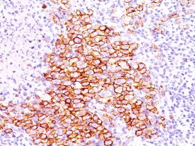
CD30(CD30/412), CF405S conjugate, 0.1mg/mL [26628-22-8]
BNC040412
ApplicationsImmunoHistoChemistry, ImmunoHistoChemistry Paraffin
Product group Antibodies
TargetTNFRSF8
Overview
- SupplierBiotium
- Product NameCD30(CD30/412), CF405S conjugate, 0.1mg/mL
- Delivery Days Customer9
- ApplicationsImmunoHistoChemistry, ImmunoHistoChemistry Paraffin
- CertificationResearch Use Only
- ClonalityMonoclonal
- Clone IDCD30/412
- Concentration0.1 mg/ml
- ConjugateOther Conjugate
- Gene ID943
- Target nameTNFRSF8
- Target descriptionTNF receptor superfamily member 8
- Target synonymsCD30; CD30L receptor; cytokine receptor CD30; D1S166E; Ki-1; Ki-1 antigen; lymphocyte activation antigen CD30; tumor necrosis factor receptor superfamily member 8
- HostMouse
- IsotypeIgG1
- Protein IDP28908
- Protein NameTumor necrosis factor receptor superfamily member 8
- Scientific DescriptionRecognizes a single chain glycoprotein of 105/120 kDa, identified as CD30/Ki-1. CD30 is synthesized as a 90 kDa precursor, which is processed in the Golgi complex into a membrane-bound phosphorylated mature 105/120 kDa glycoprotein. In Hodgkins disease, CD30/Ki-1 antigen is expressed by mononuclear-Hodgkin and multinucleated Reed-Sternberg cells. It is also expressed by the tumor cells of a majority of anaplastic large cell lymphomas as well as by a varying proportion of activated T and B cells. This MAb distinguishes large cell lymphomas derived from activated lymphoid cells from histiocytic malignancies and lymphomas derived from resting and precursor lymphoid cells or from anaplastic carcinomas. About one third of the Ki-1 positive lymphomas lack the leukocyte common antigen (CD45). Primary antibodies are available purified, or with a selection of fluorescent CF® Dyes and other labels. CF® Dyes offer exceptional brightness and photostability. Note: Conjugates of blue fluorescent dyes like CF®405S and CF®405M are not recommended for detecting low abundance targets, because blue dyes have lower fluorescence and can give higher non-specific background than other dye colors.
- SourceAnimal
- Storage Instruction2°C to 8°C
- UNSPSC12352203
Related products
Product group Antibodies
TNFRSF8 AntibodyCSB-PA172042
ApplicationsELISA, ImmunoHistoChemistry
TargetTNFRSF8
- SizePrice
Product group Antibodies
ApplicationsImmunoPrecipitation, Western Blot, ImmunoCytoChemistry, ImmunoHistoChemistry
TargetTNFRSF8
- SizePrice
Product group Antibodies
CD30 antibody [MEM-268] (FITC)GTX79953
ApplicationsFlow Cytometry
TargetTNFRSF8
- SizePrice

Product group Antibodies
Anti-CD30/TNFRSF8 Antibody Picoband(r)A01225-2-CARRIER-FREE
ApplicationsWestern Blot, ImmunoHistoChemistry
TargetTNFRSF8
- SizePrice
Product group Antibodies
ApplicationsFlow Cytometry, ImmunoFluorescence, ImmunoPrecipitation, Western Blot, ImmunoCytoChemistry, ImmunoHistoChemistry
TargetTNFRSF8
- SizePrice
Product group Antibodies
Anti-TNFRSF8 Antibody144-07651
ApplicationsWestern Blot, ImmunoHistoChemistry
TargetTNFRSF8
- SizePrice

Product group Antibodies
Anti-TNFRSF8 AntibodyAMAB91800
ApplicationsImmunoHistoChemistry
TargetTNFRSF8
- SizePrice