

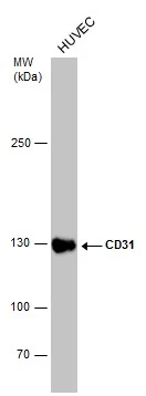
Whole cell extract (30 μg) was separated by 5% SDS-PAGE, and the membrane was blotted with CD31 antibody [N3C2], Internal (GTX110602) diluted at 1:10000.
 diluted at 1:250.
Antigen Retrieval: Trilogy? (EDTA based, pH 8.0) buffer, 15min")
![CD31 antibody [N3C2], Internal detects CD31 protein at cell membrane by immunohistochemical analysis. Sample: Paraffin-embedded human tonsil. CD31 stained by CD31 antibody [N3C2], Internal (GTX110602) diluted at 1:5000.
Antigen Retrieval: Citrate buffer, pH 6.0, 15 min](https://www.genetex.com/upload/website/prouct_img/normal/GTX110602/GTX110602_40037_20171201_IHC-P_1_w_23060500_637.webp "CD31 antibody [N3C2], Internal detects CD31 protein at cell membrane by immunohistochemical analysis. Sample: Paraffin-embedded human tonsil. CD31 stained by CD31 antibody [N3C2], Internal (GTX110602) diluted at 1:5000.
Antigen Retrieval: Citrate buffer, pH 6.0, 15 min")
Whole cell extract (30 μg) was separated by 5% SDS-PAGE, and the membrane was blotted with CD31 antibody [N3C2], Internal (GTX110602) diluted at 1:10000.
CD31 antibody [N3C2], Internal
GTX110602
ApplicationsWestern Blot, ImmunoHistoChemistry, ImmunoHistoChemistry Frozen, ImmunoHistoChemistry Paraffin
Product group Antibodies
ReactivityHuman
TargetPECAM1
Overview
- SupplierGeneTex
- Product NameCD31 antibody [N3C2], Internal
- Delivery Days Customer9
- Application Supplier NoteWB: 1:5000-1:10000. IHC-P: 1:100-1:1000. *Optimal dilutions/concentrations should be determined by the researcher.Not tested in other applications.
- ApplicationsWestern Blot, ImmunoHistoChemistry, ImmunoHistoChemistry Frozen, ImmunoHistoChemistry Paraffin
- CertificationResearch Use Only
- ClonalityPolyclonal
- Concentration1 mg/ml
- ConjugateUnconjugated
- Gene ID5175
- Target namePECAM1
- Target descriptionplatelet and endothelial cell adhesion molecule 1
- Target synonymsCD31, CD31/EndoCAM, GPIIA', PECA1, PECAM-1, endoCAM, platelet endothelial cell adhesion molecule, CD31 antigen, platelet endothelial cell adhesion molecule-1
- HostRabbit
- IsotypeIgG
- Protein IDP16284
- Protein NamePlatelet endothelial cell adhesion molecule
- Scientific DescriptionInduces susceptibility to atherosclerosis (By similarity). Cell adhesion molecule which is required for leukocyte transendothelial migration (TEM) under most inflammatory conditions. Tyr-690 plays a critical role in TEM and is required for efficient trafficking of PECAM1 to and from the lateral border recycling compartment (LBRC) and is also essential for the LBRC membrane to be targeted around migrating leukocytes. Prevents phagocyte ingestion of closely apposed viable cells by transmitting detachment signals, and changes function on apoptosis, promoting tethering of dying cells to phagocytes (the encounter of a viable cell with a phagocyte via the homophilic interaction of PECAM1 on both cell surfaces leads to the viable cells active repulsion from the phagocyte. During apoptosis, the inside-out signaling of PECAM1 is somehow disabled so that the apoptotic cell does not actively reject the phagocyte anymore. The lack of this repulsion signal together with the interaction of the eat-me signals and their respective receptors causes the attachment of the apoptotic cell to the phagocyte, thus triggering the process of engulfment). Isoform Delta15 is unable to protect against apoptosis. Modulates BDKRB2 activation. Regulates bradykinin- and hyperosmotic shock-induced ERK1/2 activation in human umbilical cord vein cells (HUVEC).
- ReactivityHuman
- Storage Instruction-20°C or -80°C,2°C to 8°C
- UNSPSC12352203
References
- He Y, Tacconi C, Dieterich LC, et al. Novel Blood Vascular Endothelial Subtype-Specific Markers in Human Skin Unearthed by Single-Cell Transcriptomic Profiling. Cells. 2022,11(7). doi: 10.3390/cells11071111Read this paper
- Feng X, Zhou L, Mao X, et al. Association of a reduction of G‑protein coupled receptor 30 expression and the pathogenesis of preeclampsia. Mol Med Rep. 2017,16(5):5997-6003. doi: 10.3892/mmr.2017.7341Read this paper
Datasheet
Related products
Product group Antibodies

Anti-CD31 [JC70]Ab01338-1.1
ApplicationsFlow Cytometry, ImmunoFluorescence, Western Blot, ImmunoHistoChemistry
ReactivityHuman, Mouse, Rabbit
TargetPECAM1
- SizePrice
Product group Antibodies

PECAM-1 (Phospho-Tyr713) AntibodyABX012447
ApplicationsImmunoFluorescence, Western Blot, ELISA, ImmunoCytoChemistry
- SizePrice
Product group Antibodies
Anti-CD31 Antibody, PE136-18092
ApplicationsFlow Cytometry
ReactivityHuman, Mouse
TargetPECAM1
- SizePrice
Product group Antibodies
CD31 antibodyGTX04325
ApplicationsFlow Cytometry, ImmunoFluorescence, Western Blot, ImmunoCytoChemistry, ImmunoHistoChemistry, ImmunoHistoChemistry Frozen, ImmunoHistoChemistry Paraffin
ReactivityBovine, Human, Mouse, Porcine, Rabbit, Rat
TargetPECAM1
- SizePrice
![IHC-P analysis of human renal cortex tissue using GTX04395 CD31 antibody [MSVA-031M] HistoMAX?. Strong CD31 staining of capillary endothelium in stroma and glomeruli of the kidney cortex.](https://www.genetex.com/upload/website/prouct_img/normal/GTX04395/GTX04395_20230728_IHC-P_20_23072722_543.webp)
Product group Antibodies
ApplicationsImmunoHistoChemistry, ImmunoHistoChemistry Paraffin
ReactivityHuman
TargetPECAM1
- SizePrice
Product group Antibodies
References
CD31 antibody [GT0010]GTX632645
ApplicationsFlow Cytometry
ReactivityHuman, Mouse
TargetPECAM1
- SizePrice
![IHC-P analysis of human tonsil tissue using GTX73514 CD31 antibody [JC/70A] (ready-to-use).](https://www.genetex.com/upload/website/prouct_img/normal/GTX73514/GTX73514_20191203_IHC-P_160_w_23061322_281.webp)
Product group Antibodies
References
ApplicationsImmunoHistoChemistry, ImmunoHistoChemistry Paraffin
ReactivityHuman, Mouse
TargetPECAM1
- SizePrice
Product group Antibodies
References
CD31 antibody [WM59] (PE)GTX75302
ApplicationsFlow Cytometry
ReactivityHuman, Monkey, Primate, Rabbit
TargetPECAM1
- SizePrice
![FACS analysis of human peripheral blood using GTX78327 CD31 antibody [MEM-05] (Biotin).](https://www.genetex.com/upload/website/prouct_img/normal/GTX78327/GTX78327_20191025_AP_006_293_w_23061322_726.webp)
Product group Antibodies
CD31 antibody [MEM-05] (Biotin)GTX78327
ApplicationsFlow Cytometry, Western Blot
ReactivityHuman
TargetPECAM1
- SizePrice