

![CD31 / PECAM-1(JC/70A), Biotin conjugate, 0.1mg/mL [26628-22-8]](https://biotium.com/wp-content/uploads/2016/12/BNUB0639-1-1.jpg "CD31 / PECAM-1(JC/70A), Biotin conjugate, 0.1mg/mL [26628-22-8]")
![CD31 / PECAM-1(JC/70A), Biotin conjugate, 0.1mg/mL [26628-22-8]](https://biotium.com/wp-content/uploads/2016/12/BNUB0639-2-1.jpg "CD31 / PECAM-1(JC/70A), Biotin conjugate, 0.1mg/mL [26628-22-8]")
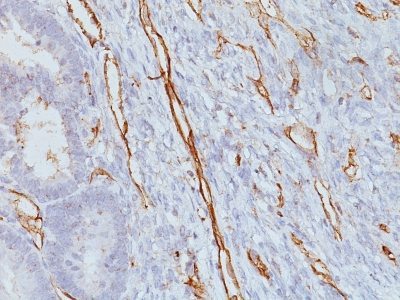
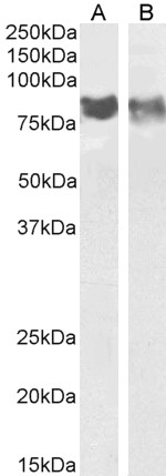
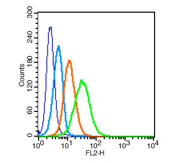
CD31 / PECAM-1(JC/70A), Biotin conjugate, 0.1mg/mL [26628-22-8]
BNCB0639
ApplicationsFlow Cytometry, Western Blot, ImmunoHistoChemistry, ImmunoHistoChemistry Paraffin
Product group Antibodies
ReactivityBovine, Human, Monkey, Mouse, Rabbit
TargetPECAM1
Overview
- SupplierBiotium
- Product NameCD31 / PECAM-1(JC/70A), Biotin conjugate, 0.1mg/mL [26628-22-8]
- Delivery Days Customer9
- ApplicationsFlow Cytometry, Western Blot, ImmunoHistoChemistry, ImmunoHistoChemistry Paraffin
- CAS Number26628-22-8
- CertificationResearch Use Only
- ClonalityMonoclonal
- Clone IDJC/70A
- Concentration0.1 mg/ml
- ConjugateBiotin
- Gene ID5175
- Target namePECAM1
- Target descriptionplatelet and endothelial cell adhesion molecule 1
- Target synonymsCD31, CD31/EndoCAM, GPIIA', PECA1, PECAM-1, endoCAM, platelet endothelial cell adhesion molecule, CD31 antigen, platelet endothelial cell adhesion molecule-1
- HostMouse
- IsotypeIgG1
- Protein IDP16284
- Protein NamePlatelet endothelial cell adhesion molecule
- Scientific DescriptionCD31 (PECAM-1) is a transmembrane glycoprotein member of the immunoglobulin supergene family of adhesion molecules. CD31 is expressed by stem cells of the hematopoietic system and is primarily used to identify and concentrate these cells for experimental studies as well as for bone marrow transplantation. Anti-CD31 has shown to be highly specific and sensitive for vascular endothelial cells. Staining of nonvascular tumors (excluding hematopoietic neoplasms) is rare. CD31 MAb reacts with normal, benign, and malignant endothelial cells which make up blood vessel lining. The level of CD31 expression can help to determine the degree of tumor angiogenesis, and a high level of CD31 expression may imply a rapidly growing tumor and potentially a predictor of tumor recurrence.Primary antibodies are available purified, or with a selection of fluorescent CF® Dyes and other labels. CF® Dyes offer exceptional brightness and photostability. Note: Conjugates of blue fluorescent dyes like CF®405S and CF®405M are not recommended for detecting low abundance targets, because blue dyes have lower fluorescence and can give higher non-specific background than other dye colors.
- SourceAnimal
- ReactivityBovine, Human, Monkey, Mouse, Rabbit
- Storage Instruction2°C to 8°C,RT
- UNSPSC41116161
MSDS
Related products
Product group Antibodies
Anti-CD31 AntibodyA286090
ApplicationsWestern Blot, ELISA
ReactivityMouse, Rat
- SizePrice
Product group Antibodies

PECAM-1 (Phospho-Tyr713) AntibodyABX012447
ApplicationsImmunoFluorescence, Western Blot, ELISA, ImmunoCytoChemistry
- SizePrice
Product group Antibodies
Anti-CD31 Antibody, PE136-18092
ApplicationsFlow Cytometry
ReactivityHuman, Mouse
TargetPECAM1
- SizePrice
Product group Antibodies

Anti-CD31 [JC70]Ab01338-1.1
ApplicationsFlow Cytometry, ImmunoFluorescence, Western Blot, ImmunoHistoChemistry
ReactivityHuman, Mouse, Rabbit
TargetPECAM1
- SizePrice
Product group Antibodies
Anti-PECAM1 AntibodyAMAB91986
ApplicationsImmunoHistoChemistry
ReactivityHuman
TargetPECAM1
- SizePrice
Product group Antibodies

ApplicationsFlow Cytometry
ReactivityMouse
TargetPECAM1
- SizePrice
Product group Antibodies
References
CD31 Polyclonal AntibodyBS-0195R
ApplicationsFlow Cytometry, ImmunoFluorescence, Western Blot, ELISA, ImmunoCytoChemistry, ImmunoHistoChemistry, ImmunoHistoChemistry Frozen, ImmunoHistoChemistry Paraffin
ReactivityBovine, Canine, Human, Porcine, Rabbit, Sheep
TargetPECAM1
- SizePrice
Product group Antibodies

ApplicationsImmunoFluorescence, ELISA, ImmunoHistoChemistry
ReactivityHuman
TargetPECAM1
- SizePrice
Product group Antibodies
Pecam1 Polyclonal AntibodyCAC07879
ApplicationsImmunoFluorescence, Western Blot, ELISA, ImmunoHistoChemistry
ReactivityMouse, Rat
TargetPECAM1
- SizePrice