

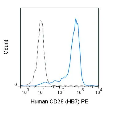
FACS analysis of human peripheral blood monocytes using GTX01499-08 CD38 antibody [HB7] (PE). Solid lone : primary antibody Dashed line : isotype control antibody amount : 0.125 μg (5 μl)
FACS analysis of human peripheral blood monocytes using GTX01499-08 CD38 antibody [HB7] (PE). Solid lone : primary antibody Dashed line : isotype control antibody amount : 0.125 μg (5 μl)
CD38 antibody [HB7] (PE)
GTX01499-08
ApplicationsFlow Cytometry
Product group Antibodies
ReactivityHuman
TargetCD38
Overview
- SupplierGeneTex
- Product NameCD38 antibody [HB7] (PE)
- Delivery Days Customer9
- Application Supplier NoteFACS: 0.125 microg (5 microl) for 105-108 cells in 100 microl sample per test. *Optimal dilutions/concentrations should be determined by the researcher.Not tested in other applications.
- ApplicationsFlow Cytometry
- CertificationResearch Use Only
- ClonalityMonoclonal
- Clone IDHB7
- Concentration0.025 mg/ml
- ConjugateRPE
- Gene ID952
- Target nameCD38
- Target descriptionCD38 molecule
- Target synonymsADPRC 1, ADPRC1, cADPR1, ADP-ribosyl cyclase/cyclic ADP-ribose hydrolase 1, 2'-phospho-ADP-ribosyl cyclase, 2'-phospho-cyclic-ADP-ribose transferase, ADP-ribosyl cyclase 1, CD38 antigen (p45), NAD(+) nucleosidase, cluster of differentiation 38, cyclic ADP-ribose hydrolase 1, ecto-nicotinamide adenine dinucleotide glycohydrolase
- HostMouse
- IsotypeIgG1
- Protein IDP28907
- Protein NameADP-ribosyl cyclase/cyclic ADP-ribose hydrolase 1
- Scientific DescriptionThe protein encoded by this gene is a non-lineage-restricted, type II transmembrane glycoprotein that synthesizes and hydrolyzes cyclic adenosine 5-diphosphate-ribose, an intracellular calcium ion mobilizing messenger. The release of soluble protein and the ability of membrane-bound protein to become internalized indicate both extracellular and intracellular functions for the protein. This protein has an N-terminal cytoplasmic tail, a single membrane-spanning domain, and a C-terminal extracellular region with four N-glycosylation sites. Crystal structure analysis demonstrates that the functional molecule is a dimer, with the central portion containing the catalytic site. It is used as a prognostic marker for patients with chronic lymphocytic leukemia. Alternative splicing results in multiple transcript variants. [provided by RefSeq, Sep 2015]
- ReactivityHuman
- Storage Instruction2°C to 8°C
- UNSPSC12352203
References
- Llinàs L, Lázaro A, de Salort J, et al. Expression profiles of novel cell surface molecules on B-cell subsets and plasma cells as analyzed by flow cytometry. Immunol Lett. 2011,134(2):113-21. doi: 10.1016/j.imlet.2010.10.009Read this paper
- Ferrero E, Malavasi F. The metamorphosis of a molecule: from soluble enzyme to the leukocyte receptor CD38. J Leukoc Biol. 1999,65(2):151-61.Read this paper
- Mehta K, Shahid U, Malavasi F. Human CD38, a cell-surface protein with multiple functions. FASEB J. 1996,10(12):1408-17.Read this paper
- Tedder TF, Clement LT, Cooper MD. Discontinuous expression of a membrane antigen (HB-7) during B lymphocyte differentiation. Tissue Antigens. 1984,24(3):140-9.Read this paper
- Jackson DG, Bell JI. Isolation of a cDNA encoding the human CD38 (T10) molecule, a cell surface glycoprotein with an unusual discontinuous pattern of expression during lymphocyte differentiation. J Immunol. 1990,144(7):2811-5.Read this paper
Datasheet
Related products
Product group Antibodies
ApplicationsFlow Cytometry
ReactivityHuman
TargetCD38
- SizePrice
Product group Antibodies

Anti-CD38 [AT.1], Mouse IgG1, kappaAB04337-1.1
ReactivityHuman
TargetCD38
- SizePrice
Product group Antibodies
Anti-Human CD38 Antibody, Purified136-00046
ApplicationsFlow Cytometry
ReactivityHuman
- SizePrice

Product group Antibodies
Anti-CD38 Antibody Picoband(r)A00193-3-CARRIER-FREE
ApplicationsFlow Cytometry, ImmunoFluorescence, Western Blot, ImmunoCytoChemistry, ImmunoHistoChemistry
ReactivityHuman
TargetCD38
- SizePrice
Product group Antibodies
CD38 antibodyGTX132579
ApplicationsWestern Blot
ReactivityHuman
TargetCD38
- SizePrice
![IHC-P analysis of human tonsil tissue section using GTX02603 CD38 antibody [CD38/4247R].](https://www.genetex.com/upload/website/prouct_img/normal/GTX02603/GTX02603_20210319_IHC-P_w_23053122_213.webp)
Product group Antibodies
References
CD38 antibody [CD38/4247R]GTX02603
ApplicationsWestern Blot, ImmunoHistoChemistry, ImmunoHistoChemistry Paraffin
ReactivityHuman
TargetCD38
- SizePrice
Product group Antibodies
CD38 antibodyGTX03746
ApplicationsImmunoFluorescence, Western Blot, ImmunoCytoChemistry, ImmunoHistoChemistry, ImmunoHistoChemistry Paraffin
ReactivityHuman, Mouse, Rat
TargetCD38
- SizePrice
![IHC-P analysis of human appendix tissue using GTX04967 CD38 antibody [MSVA-038R] HistoMAX?. In the appendix CD38 staining occurs in various inflammatory cells while the epithelial cells remain CD38 negative.](https://www.genetex.com/upload/website/prouct_img/normal/GTX04967/GTX04967_20241028_IHC-P_24102820_677.webp)
Product group Antibodies
ApplicationsImmunoHistoChemistry, ImmunoHistoChemistry Paraffin
ReactivityHuman
TargetCD38
- SizePrice