

CD3e (T-Cell Marker)(rC3e/1308), 0.2mg/mL [26628-22-8]
BNUB3631
ApplicationsImmunoHistoChemistry, ImmunoHistoChemistry Paraffin
Product group Antibodies
ReactivityHuman
TargetCD3E
Overview
- SupplierBiotium
- Product NameCD3e (T-Cell Marker)(rC3e/1308), 0.2mg/mL [26628-22-8]
- Delivery Days Customer9
- ApplicationsImmunoHistoChemistry, ImmunoHistoChemistry Paraffin
- CAS Number26628-22-8
- CertificationResearch Use Only
- ClonalityMonoclonal
- Clone IDrC3e/1308
- Concentration0.2 mg/ml
- Gene ID916
- Target nameCD3E
- Target descriptionCD3 epsilon subunit of T-cell receptor complex
- Target synonymsCD3epsilon, IMD18, T3E, TCRE, T-cell surface glycoprotein CD3 epsilon chain, CD3-epsilon, CD3e antigen, epsilon polypeptide (TiT3 complex), CD3e molecule, epsilon (CD3-TCR complex), T-cell antigen receptor complex, epsilon subunit of T3, T-cell surface antigen T3/Leu-4 epsilon chain
- HostMouse
- IsotypeIgG1
- Protein IDP07766
- Protein NameT-cell surface glycoprotein CD3 epsilon chain
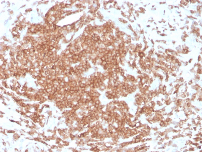
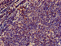
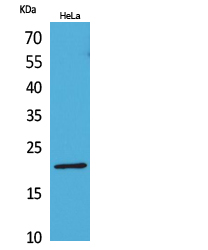
- Scientific DescriptionRecognizes the epsilon-chain of CD3, which consists of five different polypeptide chains (designated as gamma, delta, epsilon, zeta, and eta) with MW ranging from 16-28 kDa. The CD3 complex is closely associated at the lymphocyte cell surface with the T cell antigen receptor (TCR). Reportedly, CD3 complex is involved in signal transduction to the T cell interior following antigen recognition. The CD3 antigen is first detectable in early thymocytes and probably represents one of the earliest signs of commitment to the T cell lineage. In cortical thymocytes, CD3 is predominantly intra-cytoplasmic. However, in medullary thymocytes, it appears on the T cell surface. CD3 antigen is a highly specific marker for T cells, and is present in majority of T cell neoplasms. Primary antibodies are available purified, or with a selection of fluorescent CF® Dyes and other labels. CF® Dyes offer exceptional brightness and photostability. Note: Conjugates of blue fluorescent dyes like CF®405S and CF®405M are not recommended for detecting low abundance targets, because blue dyes have lower fluorescence and can give higher non-specific background than other dye colors.
- SourceAnimal
- ReactivityHuman
- Storage Instruction2°C to 8°C,RT
- UNSPSC41116161
MSDS
Related products
Product group Antibodies
CD3E AntibodyCSB-PA004931HA01HU
ApplicationsImmunoFluorescence, ELISA, ImmunoHistoChemistry
ReactivityHuman
TargetCD3E
- SizePrice
Product group Antibodies
Anti-CD3E AntibodyA101513
ApplicationsWestern Blot, ELISA
ReactivityHuman
- SizePrice
Product group Antibodies
Anti-CD3E Antibody144-01753
ApplicationsImmunoPrecipitation, Western Blot, ImmunoHistoChemistry
ReactivityHuman, Mouse, Rat
TargetCD3E
- SizePrice

Product group Antibodies
ApplicationsFlow Cytometry
ReactivityHuman
TargetCD3E
- SizePrice

Product group Antibodies
Anti-CD3E AntibodyAMAB90876
ApplicationsWestern Blot, ImmunoHistoChemistry
ReactivityHuman
TargetCD3E
- SizePrice
Product group Antibodies

Anti-CD3 epsilon [UCHT1]Ab00112-1.1
ApplicationsFlow Cytometry, ImmunoHistoChemistry
ReactivityHuman
TargetCD3E
- SizePrice

Product group Antibodies
ReactivityHuman
TargetCD3E
- SizePrice
Product group Antibodies

CD3E Antibody (clone 145-2C11, FITC)LS-C812208
ApplicationsFlow Cytometry
ReactivityMouse
TargetCD3E
- SizePrice