

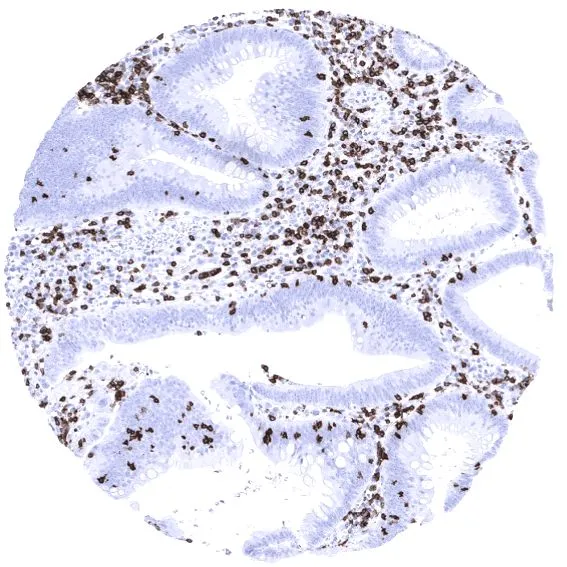
IHC-P analysis of human colorectal adenocarcinoma (COAD) tissue using GTX04390 CD7 antibody [MSVA-007R] HistoMAX?. Strong CD7 staining of a large fraction of tumor infiltrating lymphocytes in a colorectal adenocarcinoma. Tumor glands are completely CD7 negative.
![IHC-P analysis of human lung adenocarcinoma (LUAD) tissue using GTX04390 CD7 antibody [MSVA-007R] HistoMAX?. Strong membranous CD7 expression of an adenocarcinoma of the lung.](https://www.genetex.com/upload/website/prouct_img/normal/GTX04390/GTX04390_20230728_IHC-P_144_23072722_653.webp "IHC-P analysis of human lung adenocarcinoma (LUAD) tissue using GTX04390 CD7 antibody [MSVA-007R] HistoMAX?. Strong membranous CD7 expression of an adenocarcinoma of the lung.")
![IHC-P analysis of human tonsil tissue using GTX04390 CD7 antibody [MSVA-007R] HistoMAX?. Strong CD7 positivity of T lymphocytes mainly in the interfollicular area.](https://www.genetex.com/upload/website/prouct_img/normal/GTX04390/GTX04390_20230728_IHC-P_274_23072723_407.webp "IHC-P analysis of human tonsil tissue using GTX04390 CD7 antibody [MSVA-007R] HistoMAX?. Strong CD7 positivity of T lymphocytes mainly in the interfollicular area.")
IHC-P analysis of human colorectal adenocarcinoma (COAD) tissue using GTX04390 CD7 antibody [MSVA-007R] HistoMAX?. Strong CD7 staining of a large fraction of tumor infiltrating lymphocytes in a colorectal adenocarcinoma. Tumor glands are completely CD7 negative.
CD7 antibody [MSVA-007R] HistoMAX(tm)
GTX04390
ApplicationsImmunoHistoChemistry, ImmunoHistoChemistry Paraffin
Product group Antibodies
ReactivityHuman
TargetCD7
Overview
- SupplierGeneTex
- Product NameCD7 antibody [MSVA-007R] HistoMAX(tm)
- Delivery Days Customer9
- Application Supplier NoteIHC-P: 1:100-1:200. *Optimal dilutions/concentrations should be determined by the researcher.Not tested in other applications.
- ApplicationsImmunoHistoChemistry, ImmunoHistoChemistry Paraffin
- CertificationResearch Use Only
- ClonalityMonoclonal
- Clone IDMSVA-007R
- Concentration0.2 mg/ml
- ConjugateUnconjugated
- Gene ID924
- Target nameCD7
- Target descriptionCD7 molecule
- Target synonymsGP40, LEU-9, TP41, Tp40, T-cell antigen CD7, CD7 antigen (p41), T-cell leukemia antigen, T-cell surface antigen Leu-9, p41 protein
- HostRabbit
- IsotypeIgG
- Protein IDP09564
- Protein NameT-cell antigen CD7
- Scientific DescriptionThis gene encodes a transmembrane protein which is a member of the immunoglobulin superfamily. This protein is found on thymocytes and mature T cells. It plays an essential role in T-cell interactions and also in T-cell/B-cell interaction during early lymphoid development. [provided by RefSeq, Jul 2008]
- ReactivityHuman
- Storage Instruction-20°C or -80°C,2°C to 8°C
- UNSPSC41116161
Datasheet
Related products
Product group Antibodies

Anti-CD7 [3A1E]Ab00242-23.0
ApplicationsFunctional Assay, Flow Cytometry
ReactivityHuman
TargetCD7
- SizePrice
Product group Antibodies
Anti-CD7 AntibodyA101639
ApplicationsWestern Blot, ELISA
ReactivityHuman
- SizePrice

Product group Antibodies
Anti-CD7 Antibody Picoband(r)A01974-2-CARRIER-FREE
ApplicationsFlow Cytometry, Western Blot, ELISA, ImmunoCytoChemistry, ImmunoHistoChemistry
ReactivityHuman
TargetCD7
- SizePrice
Product group Antibodies
CD7 Recombinant AntibodyBSM-60231R
ApplicationsImmunoFluorescence, Western Blot, ImmunoHistoChemistry, ImmunoHistoChemistry Frozen, ImmunoHistoChemistry Paraffin
ReactivityHuman
TargetCD7
- SizePrice
Product group Antibodies

Goat anti-CD7 (aa132-146)EB11161
ApplicationsWestern Blot, ELISA
ReactivityHuman
TargetCD7
- SizePrice
Product group Antibodies
Cd7 Polyclonal AntibodyCAC08296
ApplicationsELISA, ImmunoHistoChemistry
TargetCD7
- SizePrice
Product group Antibodies
CD7 AntibodyCSB-PA004953ESR1HU
ApplicationsELISA, ImmunoHistoChemistry
ReactivityHuman
TargetCD7
- SizePrice
![FACS analysis of human peripheral blood using GTX18276 CD7 antibody [MEM-186] (FITC).](https://www.genetex.com/upload/website/prouct_img/normal/GTX18276/GTX18276_20191025_AP_006_144_w_23060620_670.webp)
Product group Antibodies
CD7 antibody [MEM-186] (FITC)GTX18276
ApplicationsFlow Cytometry
ReactivityHuman
TargetCD7
- SizePrice