


CD79a(IGA/515), Biotin conjugate, 0.1mg/mL [26628-22-8]
BNCB0515
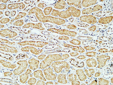
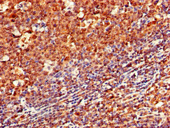
ApplicationsImmunoFluorescence, ImmunoHistoChemistry, ImmunoHistoChemistry Paraffin
Product group Antibodies
ReactivityBovine, Human, Mouse
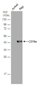
TargetCD79A
Overview
- SupplierBiotium
- Product NameCD79a(IGA/515), Biotin conjugate, 0.1mg/mL [26628-22-8]
- Delivery Days Customer9
- ApplicationsImmunoFluorescence, ImmunoHistoChemistry, ImmunoHistoChemistry Paraffin
- CAS Number26628-22-8
- CertificationResearch Use Only
- ClonalityMonoclonal
- Clone IDIGA/515
- Concentration0.1 mg/ml
- ConjugateBiotin
- Gene ID973
- Target nameCD79A
- Target descriptionCD79a molecule
- Target synonymsIGA, IGAlpha, MB-1, MB1, B-cell antigen receptor complex-associated protein alpha chain, CD79a antigen (immunoglobulin-associated alpha), CD79a molecule, immunoglobulin-associated alpha, MB-1 membrane glycoprotein, ig-alpha, membrane-bound immunoglobulin-associated protein, surface IgM-associated protein
- HostMouse
- IsotypeIgG1
- Protein IDP11912
- Protein NameB-cell antigen receptor complex-associated protein alpha chain
- Scientific DescriptionCD79 is a disulphide-linked heterodimer, consisting of mb-1 (or CD79a) and B29 (or CD79b) polypeptides, is non-covalently associated with membrane-bound immunoglobulins on B cells. This complex of mb-1 and B29 polypeptides and immunoglobulin constitute the B cell Ag receptor. CD79a first appears at pre B cell stage, early in maturation, and persists until the plasma cell stage where it is found as an intracellular component. CD79a is found in the majority of acute leukemias of precursor B cell type, in B cell lines, B cell lymphomas, and in some myelomas. It is not present in myeloid or T cell lines. Anti-CD79a is generally used to complement anti-CD20 especially for mature B-cell lymphomas after treatment with Rituximab (anti-CD20). This antibody will stain many of the same lymphomas as anti-CD20, but also is more likely to stain B-lymphoblastic lymphoma/leukemia than is anti-CD20. Anti-CD79a also stains more cases of plasma cell myeloma and occasionally some types of endothelial cells as well.Primary antibodies are available purified, or with a selection of fluorescent CF® Dyes and other labels. CF® Dyes offer exceptional brightness and photostability. Note: Conjugates of blue fluorescent dyes like CF®405S and CF®405M are not recommended for detecting low abundance targets, because blue dyes have lower fluorescence and can give higher non-specific background than other dye colors.
- SourceAnimal
- ReactivityBovine, Human, Mouse
- Storage Instruction2°C to 8°C,RT
- UNSPSC41116161
MSDS
Related products
Product group Antibodies
Anti-CD79A AntibodyA101170
ApplicationsWestern Blot, ELISA
ReactivityHuman
- SizePrice
Product group Antibodies
Anti-CD79a/IGA Antibody130-10035
ApplicationsELISA
ReactivityHuman
TargetCD79A
- SizePrice
Product group Antibodies

Anti-CD79a [HM47]Ab01318-1.1
ApplicationsImmunoPrecipitation, Western Blot, ImmunoHistoChemistry
ReactivityBovine, Chicken, Equine, Guinea Pig, Human, Mouse, Opossum, Porcine, Primate, Rabbit, Rat
TargetCD79A
- SizePrice
Product group Antibodies

ApplicationsELISA
ReactivityHuman
TargetCD79A
- SizePrice
Product group Antibodies
CD79a Monoclonal AntibodyBSM-33449M
ApplicationsImmunoFluorescence, ELISA, ImmunoHistoChemistry, ImmunoHistoChemistry Frozen, ImmunoHistoChemistry Paraffin
ReactivityHuman
TargetCD79A
- SizePrice
Product group Antibodies
ApplicationsFlow Cytometry
TargetCD79A
- SizePrice
Product group Antibodies
CD79A AntibodyCSB-PA004957LA01HU
ApplicationsImmunoFluorescence, ELISA, ImmunoHistoChemistry
ReactivityHuman
TargetCD79A
- SizePrice

Product group Antibodies
Anti-CD79A AntibodyHPA056444
ApplicationsImmunoHistoChemistry
ReactivityHuman
TargetCD79A
- SizePrice
Product group Antibodies
CD79a antibodyGTX131291
ApplicationsWestern Blot
ReactivityHuman
TargetCD79A
- SizePrice