


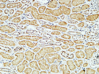
Immunohistochemical staining of human tonsil shows strong cytoplasmic positivity in germinal center cells and non-germinal center cells.
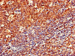
Immunohistochemical staining of human tonsil shows strong cytoplasmic positivity in germinal center cells and non-germinal center cells.
Anti-CD79A Antibody
HPA056444
ApplicationsImmunoHistoChemistry
Product group Antibodies
ReactivityHuman
TargetCD79A
Overview
- SupplierAtlas Antibodies
- Product NameAnti-CD79A Antibody
- Delivery Days Customer12
- ApplicationsImmunoHistoChemistry
- CertificationResearch Use Only
- ClonalityPolyclonal
- ConjugateUnconjugated
- Gene ID973
- Target nameCD79A
- Target descriptionCD79a molecule
- Target synonymsIGA, IGAlpha, MB-1, MB1, B-cell antigen receptor complex-associated protein alpha chain, CD79a antigen (immunoglobulin-associated alpha), CD79a molecule, immunoglobulin-associated alpha, MB-1 membrane glycoprotein, ig-alpha, membrane-bound immunoglobulin-associated protein, surface IgM-associated protein
- HostRabbit
- IsotypeIgG
- Protein IDP11912
- Protein NameB-cell antigen receptor complex-associated protein alpha chain
- Scientific DescriptionRecombinant Protein Epitope Signature Tag (PrEST) antigen sequence
- ReactivityHuman
- Storage Instruction-20°C,2°C to 8°C
- UNSPSC41116161
MSDS
Related products
Product group Antibodies
Anti-CD79A AntibodyA101170
ApplicationsWestern Blot, ELISA
ReactivityHuman
- SizePrice
Product group Antibodies
Anti-CD79a/IGA Antibody130-10035
ApplicationsELISA
ReactivityHuman
TargetCD79A
- SizePrice
Product group Antibodies

Anti-CD79a [HM47]Ab01318-1.1
ApplicationsImmunoPrecipitation, Western Blot, ImmunoHistoChemistry
ReactivityBovine, Chicken, Equine, Guinea Pig, Human, Mouse, Opossum, Porcine, Primate, Rabbit, Rat
TargetCD79A
- SizePrice
Product group Antibodies

ApplicationsELISA
ReactivityHuman
TargetCD79A
- SizePrice
Product group Antibodies
CD79a Monoclonal AntibodyBSM-33449M
ApplicationsImmunoFluorescence, ELISA, ImmunoHistoChemistry, ImmunoHistoChemistry Frozen, ImmunoHistoChemistry Paraffin
ReactivityHuman
TargetCD79A
- SizePrice
Product group Antibodies
ApplicationsFlow Cytometry
TargetCD79A
- SizePrice
Product group Antibodies
CD79A AntibodyCSB-PA004957LA01HU
ApplicationsImmunoFluorescence, ELISA, ImmunoHistoChemistry
ReactivityHuman
TargetCD79A
- SizePrice

Product group Antibodies
CD79a antibodyGTX131291
ApplicationsWestern Blot
ReactivityHuman
TargetCD79A
- SizePrice
Product group Antibodies
Anti-CD79a AntibodyCAB0331
ApplicationsImmunoFluorescence, Western Blot, ELISA, ImmunoCytoChemistry, ImmunoHistoChemistry, ImmunoHistoChemistry Paraffin
ReactivityHuman
TargetCD79A
- SizePrice