

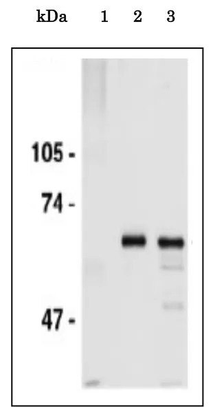
IP analysis of HeLa whole cell lysate using GTX00896 CDC6 antibody. Lane 1 : Control IP with non-immune IgG Lane 2 : Immunoprecipitates with anti-CDC6 antibody Lane 3 : Input

 using GTX00896 CDC6 antibody. Note that CDC6 abundantly localizes in nuclei of the cells at G1 and early S phase. Fixation : 4% PFA, RT, 10 min Permeabilization : ice-cold methanol, on ice, 10 min Dilution : 1:200")
")
IP analysis of HeLa whole cell lysate using GTX00896 CDC6 antibody. Lane 1 : Control IP with non-immune IgG Lane 2 : Immunoprecipitates with anti-CDC6 antibody Lane 3 : Input
CDC6 antibody
GTX00896
ApplicationsImmunoFluorescence, ImmunoPrecipitation, Western Blot, ChIP Chromatin ImmunoPrecipitation, ImmunoCytoChemistry
Product group Antibodies
ReactivityHuman, Mouse, Rat
TargetCDC6
Overview
- SupplierGeneTex
- Product NameCDC6 antibody
- Delivery Days Customer9
- Application Supplier NoteWB: 1:1000-1:3000. ICC/IF: 1:200. IP: 1:200. *Optimal dilutions/concentrations should be determined by the researcher.Not tested in other applications.
- ApplicationsImmunoFluorescence, ImmunoPrecipitation, Western Blot, ChIP Chromatin ImmunoPrecipitation, ImmunoCytoChemistry
- CertificationResearch Use Only
- ClonalityPolyclonal
- Concentration1 mg/ml
- ConjugateUnconjugated
- Gene ID990
- Target nameCDC6
- Target descriptioncell division cycle 6
- Target synonymsCDC18L, HsCDC18, HsCDC6, MGORS5, cell division control protein 6 homolog, CDC6 cell division cycle 6 homolog, CDC6-related protein, cdc18-related protein, cell division cycle 6 homolog, p62(cdc6)
- HostRabbit
- IsotypeIgG
- Protein IDQ99741
- Protein NameCell division control protein 6 homolog
- Scientific DescriptionThe protein encoded by this gene is highly similar to Saccharomyces cerevisiae Cdc6, a protein essential for the initiation of DNA replication. This protein functions as a regulator at the early steps of DNA replication. It localizes in cell nucleus during cell cyle G1, but translocates to the cytoplasm at the start of S phase. The subcellular translocation of this protein during cell cyle is regulated through its phosphorylation by Cdks. Transcription of this protein was reported to be regulated in response to mitogenic signals through transcriptional control mechanism involving E2F proteins. [provided by RefSeq, Jul 2008]
- ReactivityHuman, Mouse, Rat
- Storage Instruction-20°C or -80°C,2°C to 8°C
- UNSPSC41116161
Datasheet
Related products
Product group Antibodies
CDC6 AntibodyCSB-PA001514
ApplicationsWestern Blot, ELISA, ImmunoHistoChemistry
ReactivityHuman, Mouse
TargetCDC6
- SizePrice
Product group Antibodies
Anti-CDC6 AntibodyA101497
ApplicationsWestern Blot, ELISA
ReactivityHuman
- SizePrice
Product group Antibodies

Goat anti-CDC6EB09952
ApplicationsWestern Blot, ELISA
ReactivityHuman
TargetCDC6
- SizePrice

Product group Antibodies
Anti-CDC6 AntibodyHPA050114
ApplicationsImmunoHistoChemistry
ReactivityHuman
TargetCDC6
- SizePrice
Product group Antibodies

CDC6 Antibody (C-Terminus)LS-C368876
ApplicationsWestern Blot, ImmunoHistoChemistry, ImmunoHistoChemistry Paraffin
ReactivityHuman
TargetCDC6
- SizePrice
Product group Antibodies
CDC6 Polyclonal AntibodyCAC13465
ApplicationsELISA, ImmunoHistoChemistry
TargetCDC6
- SizePrice

Product group Antibodies
Anti-Cdc6 Antibody Picoband(r)PB9531-CARRIER-FREE
ApplicationsWestern Blot, ImmunoHistoChemistry
ReactivityHuman, Rat
TargetCDC6
- SizePrice
Product group Antibodies
CDC6 antibodyGTX108979
ApplicationsImmunoFluorescence, Western Blot, ImmunoCytoChemistry, ImmunoHistoChemistry, ImmunoHistoChemistry Paraffin
ReactivityHuman, Mouse, Rat
TargetCDC6
- SizePrice
Product group Antibodies
CDC6 (phospho Ser54) antibodyGTX55007
ApplicationsWestern Blot, ImmunoHistoChemistry, ImmunoHistoChemistry Paraffin
ReactivityHuman, Mouse, Rat
TargetCDC6
- SizePrice
Product group Antibodies
Phospho-Cdc6 (Ser54) Recombinant Antibody, Biotin ConjugatedBSM-61502R-BIOTIN
ApplicationsWestern Blot
ReactivityHuman
TargetCDC6
- SizePrice