

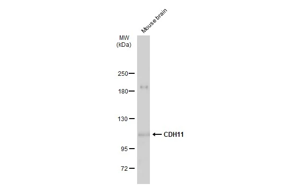
Mouse tissue extract (50 μg) was separated by 5% SDS-PAGE, and the membrane was blotted with CDH11 antibody (GTX109792) diluted at 1:1000. The HRP-conjugated anti-rabbit IgG antibody (GTX213110-01) was used to detect the primary antibody, and the signal was developed with Trident ECL plus-Enhanced.
![CDH11 antibody detects CDH11 Protein expression by immunohistochemical analysis. Sample: Frozen-sectioned adult mouse cerebellum. Green: CDH11 stained by CDH11 antibody (GTX109792) diluted at 1:250. Red: NF-H, stained by NF-H antibody [GT114] (GTX634289) diluted at 1:500. Blue: Fluoroshield with DAPI (GTX30920).
Antigen Retrieval: Citrate buffer, pH 6.0, 10 min](https://www.genetex.com/upload/website/prouct_img/normal/GTX109792/GTX109792_40051_20170831_IHC-Fr_M_w_23060500_361.webp "CDH11 antibody detects CDH11 Protein expression by immunohistochemical analysis. Sample: Frozen-sectioned adult mouse cerebellum. Green: CDH11 stained by CDH11 antibody (GTX109792) diluted at 1:250. Red: NF-H, stained by NF-H antibody [GT114] (GTX634289) diluted at 1:500. Blue: Fluoroshield with DAPI (GTX30920).
Antigen Retrieval: Citrate buffer, pH 6.0, 10 min")
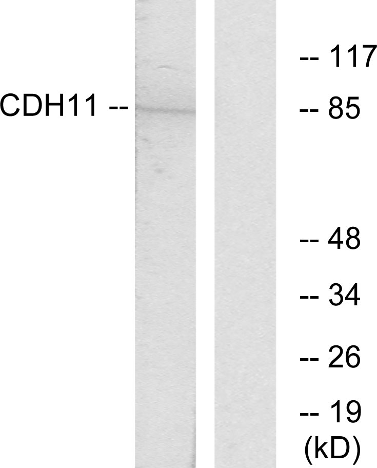
 were separated by 5% SDS-PAGE, and the membrane was blotted with CDH11 antibody (GTX109792) diluted at 1:500. The HRP-conjugated anti-rabbit IgG antibody (GTX213110-01) was used to detect the primary antibody.")
Mouse tissue extract (50 μg) was separated by 5% SDS-PAGE, and the membrane was blotted with CDH11 antibody (GTX109792) diluted at 1:1000. The HRP-conjugated anti-rabbit IgG antibody (GTX213110-01) was used to detect the primary antibody, and the signal was developed with Trident ECL plus-Enhanced.
CDH11 antibody
GTX109792
ApplicationsWestern Blot, ImmunoHistoChemistry, ImmunoHistoChemistry Frozen, ImmunoHistoChemistry Paraffin
Product group Antibodies
ReactivityHuman, Mouse
TargetCDH11
Overview
- SupplierGeneTex
- Product NameCDH11 antibody
- Delivery Days Customer9
- Application Supplier NoteWB: 1:500-1:3000. IHC-P: 1:100-1:1000. IHC-Fr: 1:100-1:1000. *Optimal dilutions/concentrations should be determined by the researcher.Not tested in other applications.
- ApplicationsWestern Blot, ImmunoHistoChemistry, ImmunoHistoChemistry Frozen, ImmunoHistoChemistry Paraffin
- CertificationResearch Use Only
- ClonalityPolyclonal
- Concentration0.5 mg/ml
- ConjugateUnconjugated
- Gene ID1009
- Target nameCDH11
- Target descriptioncadherin 11
- Target synonymsCAD11, CDHOB, ESWS, OB, OSF-4, TBHS2, cadherin-11, cadherin 11, type 2, OB-cadherin (osteoblast)
- HostRabbit
- IsotypeIgG
- Protein IDP55287
- Protein NameCadherin-11
- Scientific DescriptionThis gene encodes a type II classical cadherin from the cadherin superfamily, integral membrane proteins that mediate calcium-dependent cell-cell adhesion. Mature cadherin proteins are composed of a large N-terminal extracellular domain, a single membrane-spanning domain, and a small, highly conserved C-terminal cytoplasmic domain. Type II (atypical) cadherins are defined based on their lack of a HAV cell adhesion recognition sequence specific to type I cadherins. Expression of this particular cadherin in osteoblastic cell lines, and its upregulation during differentiation, suggests a specific function in bone development and maintenance. [provided by RefSeq]
- ReactivityHuman, Mouse
- Storage Instruction-20°C or -80°C,2°C to 8°C
- UNSPSC41116161
Datasheet
Related products
Product group Antibodies
Anti-CDH11 AntibodyA99149
ApplicationsWestern Blot, ELISA
ReactivityHuman, Mouse
- SizePrice

Product group Antibodies
Anti-OB Cadherin/CDH11 Antibody Picoband(r)A04419-1-CARRIER-FREE
ApplicationsFlow Cytometry, Western Blot, ELISA, ImmunoHistoChemistry
ReactivityHuman, Mouse, Rat
TargetCDH11
- SizePrice
Product group Antibodies
Anti-CDH11 Antibody144-08110
ApplicationsWestern Blot, ImmunoHistoChemistry
ReactivityHuman, Mouse, Rat
TargetCDH11
- SizePrice
Product group Antibodies

CDH11 / Cadherin 11 AntibodyLS-C747349
ApplicationsWestern Blot
ReactivityHuman
TargetCDH11
- SizePrice
Product group Antibodies
ApplicationsWestern Blot
ReactivityBovine, Canine, Equine, Human, Mouse, Porcine, Rabbit, Rat
TargetCDH11
- SizePrice
Product group Antibodies

Goat anti-CDH11 (aa 26-37)EB10071
ApplicationsWestern Blot, ELISA
ReactivityBovine, Canine, Human, Mouse, Rat
TargetCDH11
- SizePrice
Product group Antibodies
Cdh11 Polyclonal AntibodyCAC08301
ApplicationsImmunoFluorescence, ELISA, ImmunoHistoChemistry
TargetCDH11
- SizePrice
Product group Antibodies
CDH11 AntibodyCSB-PA005036EA01HU
ApplicationsImmunoFluorescence, ELISA, ImmunoHistoChemistry
ReactivityHuman
TargetCDH11
- SizePrice
Product group Antibodies

ApplicationsWestern Blot, ImmunoCytoChemistry, ImmunoHistoChemistry, ImmunoHistoChemistry Frozen
ReactivityHuman, Rat
TargetCDH11
- SizePrice
Product group Antibodies
CDH11 antibodyGTX04544
ApplicationsFlow Cytometry, Western Blot, ELISA, ImmunoHistoChemistry, ImmunoHistoChemistry Paraffin
ReactivityHuman, Mouse, Rat
TargetCDH11
- SizePrice