

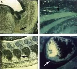
ICC staining of mouse tissue using anti-cdk9(PITALRE). The staining shows the location of mcdk9/PITALRE protein in developing mouse tissue. Arrows indicate areas of high expression. Panel A: Peroxidase-DAB immunostaining of mcdk9/PITALRE protein in the developing mouse brain in the differentiated region of the medulla oblongata just below the fourth ventricle. Similar staining is shown in Panel B in the dorsal root ganglia. Panel C: Fluorescein immunofluorescence of mcdk9IPITALRE in skeletal muscle. Similar staining is shown in Panel D in cardiac muscle. Sections from each specimen were cut at 5-7 μm, mounted on glass and dried overnight at 37oC. All sections then were deparaffinized in xylene, rehydrated through a graded alcohol series and washed in phosphate-buffered saline (PBS). PBS was used for all subsequent washes and for antiserum dilution. Tissue sections were quenched sequentially in 0.5% hydrogen peroxide and blocked with diluted 10% normal goat anti-rabbit serum. Slides were incubated at 20o C for 1 h with rabbit anti-cdk9 (GTX26544)(1:500) dilution, washed, and then reacted with diluted goat anti-rabbit biotinylated antibody for 30 min. All the slides were then reacted with streptavidin-peroxidase conjugate for 30 min at 20o C. Diaminobenzidine was used as the final chromogen and hematoxylin was used as the nuclear counterstain. Negative controls for each tissue section were prepared by substituting the primary antiserum with pre-immune serum.
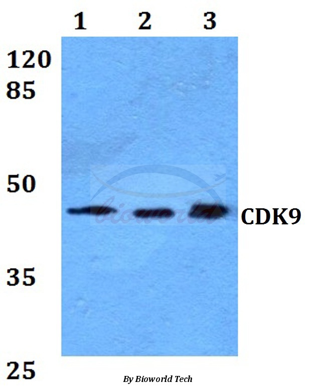
 was used for Western blot analysis of 1) PC3 and 2) DU145 prostate cancer cells (50ug per lane). Bands at the expected MW of 55 and 42 Kda were detected.")

 Lane 3 : PC-3 whole cell lysate (20 μg) Lane 4 : Null Dilution : 1:1000")
ICC staining of mouse tissue using anti-cdk9(PITALRE). The staining shows the location of mcdk9/PITALRE protein in developing mouse tissue. Arrows indicate areas of high expression. Panel A: Peroxidase-DAB immunostaining of mcdk9/PITALRE protein in the developing mouse brain in the differentiated region of the medulla oblongata just below the fourth ventricle. Similar staining is shown in Panel B in the dorsal root ganglia. Panel C: Fluorescein immunofluorescence of mcdk9IPITALRE in skeletal muscle. Similar staining is shown in Panel D in cardiac muscle. Sections from each specimen were cut at 5-7 μm, mounted on glass and dried overnight at 37oC. All sections then were deparaffinized in xylene, rehydrated through a graded alcohol series and washed in phosphate-buffered saline (PBS). PBS was used for all subsequent washes and for antiserum dilution. Tissue sections were quenched sequentially in 0.5% hydrogen peroxide and blocked with diluted 10% normal goat anti-rabbit serum. Slides were incubated at 20o C for 1 h with rabbit anti-cdk9 (GTX26544)(1:500) dilution, washed, and then reacted with diluted goat anti-rabbit biotinylated antibody for 30 min. All the slides were then reacted with streptavidin-peroxidase conjugate for 30 min at 20o C. Diaminobenzidine was used as the final chromogen and hematoxylin was used as the nuclear counterstain. Negative controls for each tissue section were prepared by substituting the primary antiserum with pre-immune serum.
CDK9 antibody
GTX26544
ApplicationsImmunoPrecipitation, Western Blot, ELISA, ImmunoHistoChemistry, ImmunoHistoChemistry Paraffin
Product group Antibodies
ReactivityHuman, Mouse
TargetCDK9
Overview
- SupplierGeneTex
- Product NameCDK9 antibody
- Delivery Days Customer9
- Application Supplier NoteWB: 1:500-1:3000. IHC-P: 1:200-1:1000. IP: 1:100. ELISA: 1:10000-1:50000. *Optimal dilutions/concentrations should be determined by the researcher.Not tested in other applications.
- ApplicationsImmunoPrecipitation, Western Blot, ELISA, ImmunoHistoChemistry, ImmunoHistoChemistry Paraffin
- CertificationResearch Use Only
- ClonalityPolyclonal
- Concentration75 mg/ml
- ConjugateUnconjugated
- Gene ID1025
- Target nameCDK9
- Target descriptioncyclin dependent kinase 9
- Target synonymsC-2k, CDC2L4, CTK1, PITALRE, TAK, cyclin-dependent kinase 9, CDC2-related kinase, cell division cycle 2-like protein kinase 4, cell division protein kinase 9, serine/threonine protein kinase PITALRE, tat-associated kinase complex catalytic subunit
- HostRabbit
- IsotypeIgG
- Protein IDP50750
- Protein NameCyclin-dependent kinase 9
- Scientific DescriptionCDK9 (PITALRE) (also known as cyclin-dependent kinase 9, Serine/threonine-protein kinase PITALRE, C-2K and Cell division cycle 2-like protein kinase 4) is a member of the cyclin-dependent protein kinase (CDK) family. CDK family members are highly similar to the gene products of S. cerevisiae cdc28, and S. pombe cdc2, and known as important cell cycle regulators. CDK9 (PITALRE) interacts with a conserved domain in the TRAF-C region of the tumor necrosis factor signal transducer TRAF2. This kinase also was found to be a component of the multiprotein complex TAK/P-TEFb, which is an elongation factor for RNA polymerase II-directed transcription and functions by phosphorylating the C-terminal domain of the largest subunit of RNA polymerase II. This protein forms a complex with and is regulated by its regulatory subunit cyclin T or cyclin K. HIV-1 Tat protein was found to interact with this protein and cyclin T, which suggested a possible involvement of this protein in AIDS. Tat stimulates human HIV-1 viral transcription elongation. This suggests that cyclin T1/cdk9(PITALRE) is one of the HIV-1 required host cellular cofactors generated during T cell activation. Cyclin T1/cdk9(PITALRE) is shown to interact with Tat to restore Tat activation in HeLa nuclear extracts depleted of P-TEFb. The cdk9(PITALRE) activity and cyclin T1 are essential for activation of transcription when tethered to the heterologous Rev response element RNA via the regulator of expression of virion Rev. CDK9 (PITALRE) is a ubiquitously expressed nuclear protein.
- ReactivityHuman, Mouse
- Storage Instruction-20°C or -80°C,2°C to 8°C
- UNSPSC41116161
Datasheet
Related products
Product group Antibodies
CDK9 AntibodyCSB-PA005079ESR1HU
ApplicationsELISA, ImmunoHistoChemistry
ReactivityHuman
TargetCDK9
- SizePrice
Product group Antibodies
Anti-CDK9 AntibodyA28226
ApplicationsWestern Blot
ReactivityHuman, Mouse, Rat
- SizePrice
Product group Antibodies

CDK9 AntibodyLS-C761129
ApplicationsWestern Blot
ReactivityBovine, Chicken, Human, Mouse, Porcine, Rat
TargetCDK9
- SizePrice

Product group Antibodies
Anti-CDK9 AntibodyHPA006738
ApplicationsChIP Chromatin ImmunoPrecipitation, ImmunoCytoChemistry, ImmunoHistoChemistry
ReactivityHuman
TargetCDK9
- SizePrice
Product group Antibodies

Goat anti-CDK9EB13048
ApplicationsWestern Blot, ELISA
ReactivityBovine, Canine, Human, Mouse, Porcine, Rat
TargetCDK9
- SizePrice
Product group Antibodies
ApplicationsImmunoPrecipitation, Western Blot, ImmunoCytoChemistry, ImmunoHistoChemistry
ReactivityMouse, Rat
TargetCDK9
- SizePrice

Product group Antibodies
Anti-Cdk9 Antibody Picoband(r)PB9537-CARRIER-FREE
ApplicationsWestern Blot, ImmunoHistoChemistry
ReactivityHuman, Rat
TargetCDK9
- SizePrice
![CDK9 antibody [N3C3] detects CDK9 protein at nucleus by immunofluorescent analysis. Sample: HeLa cells were fixed in 4% paraformaldehyde at RT for 15 min. Green: CDK9 protein stained by CDK9 antibody [N3C3] (GTX107758) diluted at 1:500. Red: phalloidin, a cytoskeleton marker, diluted at 1:200. Scale bar = 10 μm.](https://www.genetex.com/upload/website/prouct_img/normal/GTX107758/GTX107758_40688_20160120_IFA_w_23060120_269.webp)
Product group Antibodies
CDK9 antibody [N3C3]GTX107758
ApplicationsImmunoFluorescence, Western Blot, ImmunoCytoChemistry, ImmunoHistoChemistry, ImmunoHistoChemistry Paraffin
ReactivityHuman, Mouse, Rat
TargetCDK9
- SizePrice