

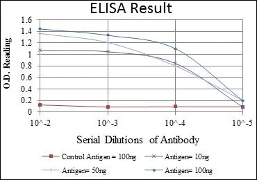
ELISA analysis of antigen using GTX60388 CEA antibody [3G12].
Red : Control antigen 100ng
Purple : Antigen 10ng
Green : Antigen 50ng
Blue : Antigen 100ng
![ICC/IF analysis of PANC-1 cells using GTX60388 CEA antibody [3G12]. Green : CEA Blue: DRAQ5 fluorescent DNA dye Red: Actin filaments](https://www.genetex.com/upload/website/prouct_img/normal/GTX60388/GTX60388_20170912_ICCIF_w_23061123_370.webp "ICC/IF analysis of PANC-1 cells using GTX60388 CEA antibody [3G12]. Green : CEA Blue: DRAQ5 fluorescent DNA dye Red: Actin filaments")
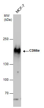
![WB analysis of HEK293 (1) and CEA(AA: 460-600)-hIgGFc transfected HEK293 (2) cell lysate using GTX60388 CEA antibody [3G12].](https://www.genetex.com/upload/website/prouct_img/normal/GTX60388/GTX60388_20170912_WB_w_23061123_283.webp "WB analysis of HEK293 (1) and CEA(AA: 460-600)-hIgGFc transfected HEK293 (2) cell lysate using GTX60388 CEA antibody [3G12].")
ELISA analysis of antigen using GTX60388 CEA antibody [3G12].
Red : Control antigen 100ng
Purple : Antigen 10ng
Green : Antigen 50ng
Blue : Antigen 100ng
CD66e antibody [3G12]
GTX60388
ApplicationsImmunoFluorescence, Western Blot, ELISA, ImmunoCytoChemistry
Product group Antibodies
ReactivityHuman
TargetCEACAM5
Overview
- SupplierGeneTex
- Product NameCD66e antibody [3G12]
- Delivery Days Customer9
- Application Supplier NoteWB: 1/500 - 1/2000. ICC/IF: 1/200 - 1/1000. ELISA: 1/10000. *Optimal dilutions/concentrations should be determined by the researcher.Not tested in other applications.
- ApplicationsImmunoFluorescence, Western Blot, ELISA, ImmunoCytoChemistry
- CertificationResearch Use Only
- ClonalityMonoclonal
- Clone ID3G12
- ConjugateUnconjugated
- Gene ID1048
- Target nameCEACAM5
- Target descriptionCEA cell adhesion molecule 5
- Target synonymsCD66e, CEA, cell adhesion molecule CEACAM5, carcinoembryonic antigen related cell adhesion molecule 5, meconium antigen 100
- HostMouse
- IsotypeIgG1
- Protein IDP06731
- Protein NameCell adhesion molecule CEACAM5
- Scientific DescriptionThis gene encodes a cell surface glycoprotein that represents the founding member of the carcinoembryonic antigen (CEA) family of proteins. The encoded protein is used as a clinical biomarker for gastrointestinal cancers and may promote tumor development through its role as a cell adhesion molecule. Additionally, the encoded protein may regulate differentiation, apoptosis, and cell polarity. This gene is present in a CEA family gene cluster on chromosome 19. Alternative splicing results in multiple transcript variants. [provided by RefSeq, Jul 2015]
- ReactivityHuman
- Storage Instruction-20°C or -80°C,2°C to 8°C
- UNSPSC12352203
Datasheet
Related products
Product group Antibodies

Anti-CEA [EB-011]Ab01002-1.1
ApplicationsELISA
ReactivityHuman
TargetCEACAM5
- SizePrice
Product group Antibodies
Anti-CEACAM5 Antibody144-60045
ApplicationsWestern Blot
ReactivityHuman, Mouse
TargetCEACAM5
- SizePrice

Product group Antibodies
Anti-CEA/CEACAM5 Antibody Picoband(r)A00356-2-CARRIER-FREE
ApplicationsFlow Cytometry, Western Blot, ELISA
ReactivityHuman
TargetCEACAM5
- SizePrice
Product group Antibodies
CD66e antibody [12-140-10]GTX01863
ApplicationsImmunoHistoChemistry, ImmunoHistoChemistry Frozen, ImmunoHistoChemistry Paraffin
ReactivityHuman
TargetCEACAM5
- SizePrice
![IHC-P analysis of human colon tissue using GTX04399 CD66e antibody [MSVA-465R] HistoMAX?. Moderate to strong CD66e immunostaining in the epithelial cells of the colon. Inflammatory and smooth muscle cells remain negative.](https://www.genetex.com/upload/website/prouct_img/normal/GTX04399/GTX04399_20230728_IHC-P_25_23072722_786.webp)
Product group Antibodies
ApplicationsImmunoHistoChemistry, ImmunoHistoChemistry Paraffin
ReactivityHuman
TargetCEACAM5
- SizePrice
Product group Antibodies
References
CD66e antibodyGTX100903
ApplicationsImmunoFluorescence, Western Blot, ImmunoCytoChemistry
ReactivityHuman
TargetCEACAM5
- SizePrice
Product group Antibodies
References
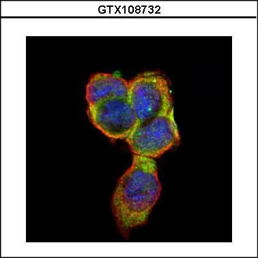
CD66e antibodyGTX108732
ApplicationsImmunoFluorescence, Western Blot, ImmunoCytoChemistry, ImmunoHistoChemistry, ImmunoHistoChemistry Paraffin
ReactivityHuman
TargetCEACAM5
- SizePrice
Product group Antibodies
CD66e antibody [N1N3]GTX113161
ApplicationsWestern Blot, ImmunoHistoChemistry, ImmunoHistoChemistry Paraffin
ReactivityHuman
TargetCEACAM5
- SizePrice