

CEP68 Antibody
CSB-PA765741LA01HU
ApplicationsELISA
Product group Antibodies
ReactivityHuman
TargetCEP68
Overview
- SupplierCusabio
- Product NameCEP68 Antibody
- Delivery Days Customer20
- ApplicationsELISA
- CertificationResearch Use Only
- ClonalityPolyclonal
- ConjugateUnconjugated
- Gene ID23177
- Target nameCEP68
- Target descriptioncentrosomal protein 68
- Target synonymsKIAA0582, centrosomal protein of 68 kDa, centrosomal protein 68kDa
- HostRabbit
- IsotypeIgG
- Protein IDQ76N32
- Protein NameCentrosomal protein of 68 kDa
- Scientific DescriptionInvolved in maintenance of centrosome cohesion, probably as part of a linker structure which prevents centrosome splitting (PubMed:18042621). Required for localization of CDK5RAP2 to the centrosome during interphase (PubMed:24554434, PubMed:25503564).
- ReactivityHuman
- Storage Instruction-20°C or -80°C
- UNSPSC41116161
Related products
Product group Antibodies
Anti-CEP68 Antibody Picoband(r)A01704-1-CARRIER-FREE
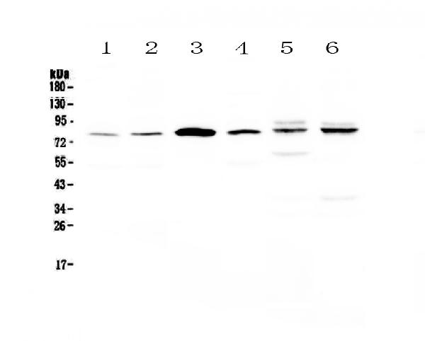
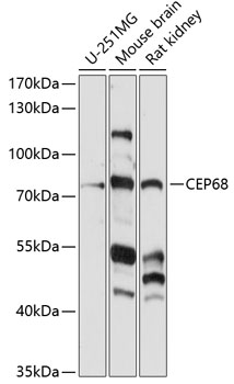
ApplicationsWestern Blot
ReactivityHuman, Mouse, Rat
TargetCEP68
- SizePrice
Product group Antibodies
Anti-CEP68 Antibody144-63475
ApplicationsWestern Blot
ReactivityHuman, Mouse, Rat
TargetCEP68
- SizePrice

Product group Antibodies
Anti-CEP68 AntibodyHPA040493
ApplicationsWestern Blot, ImmunoCytoChemistry, ImmunoHistoChemistry
ReactivityHuman
TargetCEP68
- SizePrice
Product group Antibodies

CEP68 AntibodyLS-C749841
ApplicationsWestern Blot
ReactivityHuman, Mouse, Rat
TargetCEP68
- SizePrice
Product group Antibodies
Anti-CEP68 AntibodyA91168
ApplicationsWestern Blot
ReactivityHuman, Mouse, Rat
- SizePrice