

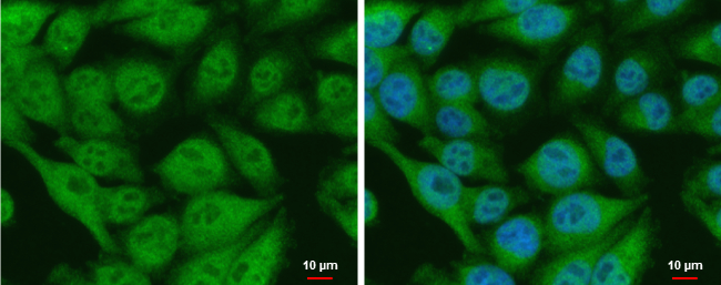
CIAPIN1 antibody detects CIAPIN1 protein at nucleus and cytoplasm by immunofluorescent analysis. Sample: A375 cells were fixed in 4% paraformaldehyde at RT for 15 min. Green: CIAPIN1 protein stained by CIAPIN1 antibody (GTX111212) diluted at 1:500. Blue: Hoechst 33342 staining. Scale bar = 10 μm.
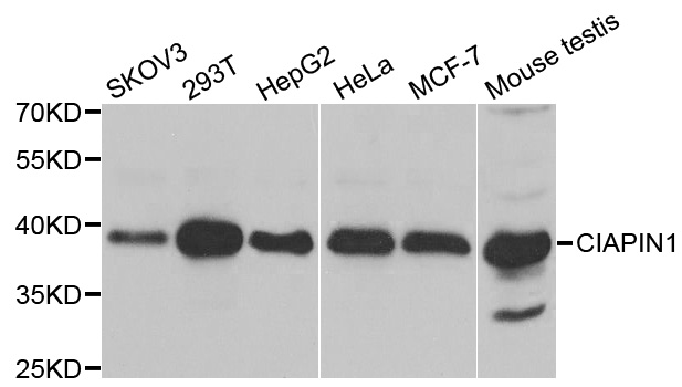
 dilution: 1:500.
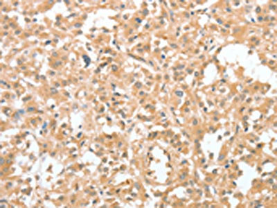
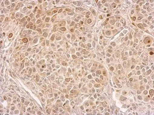
Antigen Retrieval: Trilogy? (EDTA based, pH 8.0) buffer, 15min")
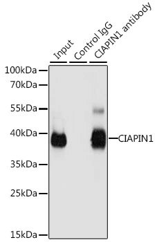
 A: Jurkat B: Raji C: K562 D: THP-1 E: HL-60 F: NCI-H929 10% SDS PAGE GTX111212 diluted at 1:1000")
 A: NIH-3T3 B: JC C: BCL-1 10% SDS PAGE GTX111212 diluted at 1:3000")
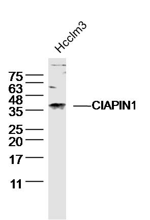
 diluted at 1:500.
Antigen Retrieval: Citrate buffer, pH 6.0, 15 min")
CIAPIN1 antibody detects CIAPIN1 protein at nucleus and cytoplasm by immunofluorescent analysis. Sample: A375 cells were fixed in 4% paraformaldehyde at RT for 15 min. Green: CIAPIN1 protein stained by CIAPIN1 antibody (GTX111212) diluted at 1:500. Blue: Hoechst 33342 staining. Scale bar = 10 μm.
CIAPIN1 antibody
GTX111212
ApplicationsImmunoFluorescence, Western Blot, ImmunoCytoChemistry, ImmunoHistoChemistry, ImmunoHistoChemistry Paraffin
Product group Antibodies
ReactivityHuman, Mouse, Rat
TargetCIAPIN1
Overview
- SupplierGeneTex
- Product NameCIAPIN1 antibody
- Delivery Days Customer9
- Application Supplier NoteWB: 1:500-1:3000. ICC/IF: 1:100-1:1000. IHC-P: 1:100-1:1000. *Optimal dilutions/concentrations should be determined by the researcher.Not tested in other applications.
- ApplicationsImmunoFluorescence, Western Blot, ImmunoCytoChemistry, ImmunoHistoChemistry, ImmunoHistoChemistry Paraffin
- CertificationResearch Use Only
- ClonalityPolyclonal
- Concentration1 mg/ml
- ConjugateUnconjugated
- Gene ID57019
- Target nameCIAPIN1
- Target descriptioncytokine induced apoptosis inhibitor 1
- Target synonymsAnamorsin, CIAE2, DRE2, PRO0915, anamorsin, fe-S cluster assembly protein DRE2 homolog, predicted protein of HQ0915
- HostRabbit
- IsotypeIgG
- Protein IDQ6FI81
- Protein NameAnamorsin
- Scientific DescriptionCIAPIN1 is a cytokine-induced inhibitor of apoptosis with no relation to apoptosis regulatory molecules of the BCL2 (MIM 151430) or CASP (see MIM 147678) families. Expression of CIAPIN1 is dependent on growth factor stimulation (Shibayama et al., 2004 [PubMed 14970183]).[supplied by OMIM]
- ReactivityHuman, Mouse, Rat
- Storage Instruction-20°C or -80°C,2°C to 8°C
- UNSPSC41116161
Datasheet
Related products
Product group Antibodies
Anti-CIAPIN1 AntibodyA31308
ApplicationsWestern Blot, ImmunoHistoChemistry
ReactivityHuman, Mouse
- SizePrice
Product group Antibodies
Anti-CIAPIN1 Antibody144-06336
ApplicationsImmunoFluorescence, ImmunoPrecipitation, Western Blot, ImmunoHistoChemistry
ReactivityHuman, Mouse
TargetCIAPIN1
- SizePrice
Product group Antibodies
CIAPIN1 Polyclonal AntibodyBS-5764R
ApplicationsImmunoFluorescence, Western Blot, ELISA, ImmunoCytoChemistry, ImmunoHistoChemistry, ImmunoHistoChemistry Frozen, ImmunoHistoChemistry Paraffin
ReactivityBovine, Human, Mouse, Porcine, Rat
TargetCIAPIN1
- SizePrice
Product group Antibodies
CIAPIN1 AntibodyCSB-PA102580
ApplicationsWestern Blot, ELISA, ImmunoHistoChemistry
ReactivityHuman
TargetCIAPIN1
- SizePrice
Product group Antibodies

CIAPIN1 / Anamorsin AntibodyLS-C401529
ApplicationsELISA, ImmunoHistoChemistry
ReactivityHuman
TargetCIAPIN1
- SizePrice

Product group Antibodies
Anti-CIAPIN1 AntibodyHPA042182
ApplicationsWestern Blot, ImmunoHistoChemistry
ReactivityHuman, Mouse, Rat
TargetCIAPIN1
- SizePrice
Product group Antibodies
CIAPIN1 antibodyGTX113988
ApplicationsImmunoFluorescence, Western Blot, ImmunoCytoChemistry, ImmunoHistoChemistry, ImmunoHistoChemistry Paraffin
ReactivityHuman
TargetCIAPIN1
- SizePrice
Product group Antibodies
CIAPIN1 antibodyGTX55572
ApplicationsImmunoFluorescence, ImmunoPrecipitation, Western Blot, ImmunoCytoChemistry, ImmunoHistoChemistry, ImmunoHistoChemistry Paraffin
ReactivityHuman, Mouse
TargetCIAPIN1
- SizePrice
Product group Antibodies
Anti-CIAPIN1 AntibodyCAB6336
ApplicationsImmunoFluorescence, ImmunoPrecipitation, Western Blot, ELISA, ImmunoCytoChemistry
ReactivityHuman
TargetCIAPIN1
- SizePrice