

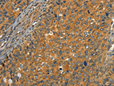
The image on the left is immunohistochemistry of paraffin-embedded Human cervical cancer tissue using CSB-PA103488(CIB1 Antibody) at dilution 1/30, on the right is treated with synthetic peptide. (Original magnification: x200)
 at dilution 1/30, on the right is treated with synthetic peptide. (Original magnification: x200)")
The image on the left is immunohistochemistry of paraffin-embedded Human cervical cancer tissue using CSB-PA103488(CIB1 Antibody) at dilution 1/30, on the right is treated with synthetic peptide. (Original magnification: x200)
CIB1 Antibody
CSB-PA103488
ApplicationsELISA, ImmunoHistoChemistry
Product group Antibodies
ReactivityHuman, Mouse, Rat
TargetCIB1
Overview
- SupplierCusabio
- Product NameCIB1 Antibody
- Delivery Days Customer20
- ApplicationsELISA, ImmunoHistoChemistry
- CertificationResearch Use Only
- ClonalityPolyclonal
- ConjugateUnconjugated
- Gene ID10519
- Target nameCIB1
- Target descriptioncalcium and integrin binding 1
- Target synonymsCIB, CIBP, EV3, KIP1, PRKDCIP, SIP2-28, calcium and integrin-binding protein 1, DNA-PK interaction protein, DNA-PKcs-interacting protein, DNA-dependent protein kinase interacting protein, SNK-interacting protein 2-28, calcium and integrin binding 1 (calmyrin), testicular secretory protein Li 9
- HostRabbit
- IsotypeIgG
- Protein IDQ99828
- Protein NameCalcium and integrin-binding protein 1
- Scientific DescriptionThis gene encodes a member of the EF-hand domain-containing calcium-binding superfamily. The encoded protein interacts with many other proteins, including the platelet integrin alpha-IIb-beta-3, DNA-dependent protein kinase, presenilin-2, focal adhesion kinase, p21 activated kinase, and protein kinase D. The encoded protein may be involved in cell survival and proliferation, and is associated with several disease states including cancer and Alzheimers disease. Alternative splicing results in multiple transcript variants.
- ReactivityHuman, Mouse, Rat
- Storage Instruction-20°C or -80°C
- UNSPSC41116161
Related products
Product group Antibodies
Anti-CIB1 AntibodyA28256
ApplicationsWestern Blot
ReactivityHuman, Rat
- SizePrice
Product group Antibodies
Anti-CIB1 Antibody144-04430
ApplicationsWestern Blot, ImmunoHistoChemistry
ReactivityHuman, Mouse, Rat
TargetCIB1
- SizePrice
Product group Antibodies
ApplicationsImmunoPrecipitation, Western Blot, ImmunoCytoChemistry, ImmunoHistoChemistry
ReactivityMouse, Rat
TargetCIB1
- SizePrice
Product group Antibodies
CIB1 Polyclonal AntibodyBS-1682R
ApplicationsImmunoFluorescence, ELISA, ImmunoCytoChemistry, ImmunoHistoChemistry, ImmunoHistoChemistry Frozen, ImmunoHistoChemistry Paraffin
ReactivityBovine, Canine, Equine, Guinea Pig, Human, Mouse, Porcine, Rabbit, Rat
TargetCIB1
- SizePrice
Product group Antibodies

CIB1 / KIP AntibodyLS-C403065
ApplicationsELISA, ImmunoHistoChemistry
ReactivityHuman, Mouse, Rat
TargetCIB1
- SizePrice
Product group Antibodies
CIB1 antibodyGTX100849
ApplicationsWestern Blot, ImmunoHistoChemistry, ImmunoHistoChemistry Paraffin
ReactivityHuman
TargetCIB1
- SizePrice

Product group Antibodies
Anti-CIB1 AntibodyHPA048825
ApplicationsImmunoCytoChemistry
ReactivityHuman
TargetCIB1
- SizePrice

Product group Antibodies
Anti-CIB1 Antibody Picoband(r)PB9489-CARRIER-FREE
ApplicationsWestern Blot
ReactivityHuman, Mouse, Rat
TargetCIB1
- SizePrice
Product group Antibodies
ApplicationsWestern Blot, ELISA
ReactivityHuman
TargetCIB1
- SizePrice