

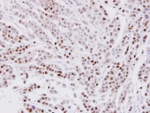
Immunohistochemical analysis of paraffin-embedded MDAMB-468 xenograft, using CIP29(GTX106633) antibody at 1:100 dilution.
Antigen Retrieval: Trilogy? (EDTA based, pH 8.0) buffer, 15min
![CIP29 antibody [N1C3] detects CIP29 protein at nucleus by immunofluorescent analysis. Sample: HeLa cells were fixed in 4% paraformaldehyde at RT for 15 min. Green: CIP29 protein stained by CIP29 antibody [N1C3] (GTX106633) diluted at 1:1000. Blue: Hoechst 33342 staining.](https://www.genetex.com/upload/website/prouct_img/normal/GTX106633/GTX106633_41017_IFA_w_23060120_945.webp "CIP29 antibody [N1C3] detects CIP29 protein at nucleus by immunofluorescent analysis. Sample: HeLa cells were fixed in 4% paraformaldehyde at RT for 15 min. Green: CIP29 protein stained by CIP29 antibody [N1C3] (GTX106633) diluted at 1:1000. Blue: Hoechst 33342 staining.")
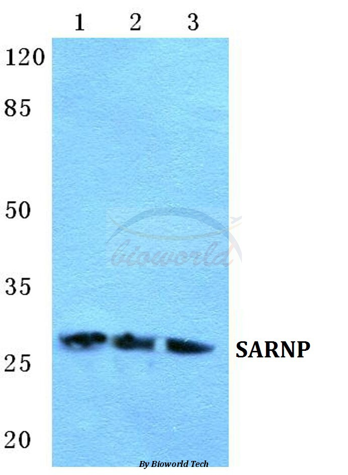
 A:293T B:A431(GTX27909) C:H1299 12% SDS PAGE GTX106633 diluted at 1:1000")
Immunohistochemical analysis of paraffin-embedded MDAMB-468 xenograft, using CIP29(GTX106633) antibody at 1:100 dilution.
Antigen Retrieval: Trilogy? (EDTA based, pH 8.0) buffer, 15min
CIP29 antibody [N1C3]
GTX106633
ApplicationsImmunoFluorescence, Western Blot, ImmunoCytoChemistry, ImmunoHistoChemistry, ImmunoHistoChemistry Paraffin
Product group Antibodies
ReactivityHuman
TargetSARNP
Overview
- SupplierGeneTex
- Product NameCIP29 antibody [N1C3]
- Delivery Days Customer9
- Application Supplier NoteWB: 1:500-1:3000. ICC/IF: 1:100-1:1000. IHC-P: 1:100-1:1000. *Optimal dilutions/concentrations should be determined by the researcher.Not tested in other applications.
- ApplicationsImmunoFluorescence, Western Blot, ImmunoCytoChemistry, ImmunoHistoChemistry, ImmunoHistoChemistry Paraffin
- CertificationResearch Use Only
- ClonalityPolyclonal
- Concentration0.33 mg/ml
- ConjugateUnconjugated
- Gene ID84324
- Target nameSARNP
- Target descriptionSAP domain containing ribonucleoprotein
- Target synonymsCIP29, HCC1, HSPC316, THO1, SAP domain-containing ribonucleoprotein, cytokine induced protein 29 kDa, cytokine-induced protein of 29 kDa, hepatocellular carcinoma 1, nuclear protein Hcc-1, proliferation associated cytokine-inducible protein CIP29
- HostRabbit
- IsotypeIgG
- Protein IDP82979
- Protein NameSAP domain-containing ribonucleoprotein
- Scientific DescriptionThis gene encodes a protein that is upregulated in response to various cytokines. The encoded protein may play a role in cell cycle progression. A translocation between this gene and the myeloid/lymphoid leukemia gene, resulting in expression of a chimeric protein, has been associated with acute myelomonocytic leukemia. Pseudogenes exist on chromosomes 7 and 8. Alternatively spliced transcript variants have been described. [provided by RefSeq]
- ReactivityHuman
- Storage Instruction-20°C or -80°C,2°C to 8°C
- UNSPSC41116161
Datasheet
Related products
Product group Antibodies
SARNP AntibodyCSB-PA070014
ApplicationsWestern Blot, ELISA, ImmunoHistoChemistry
ReactivityHuman, Mouse, Rat
TargetSARNP
- SizePrice

Product group Antibodies
Anti-HCC1 SARNP AntibodyA11119
ApplicationsImmunoFluorescence, Western Blot, ELISA, ImmunoCytoChemistry, ImmunoHistoChemistry
ReactivityHuman, Mouse, Rat
TargetSARNP
- SizePrice
Product group Antibodies
Anti-SARNP AntibodyA27923
ApplicationsWestern Blot, ImmunoCytoChemistry
ReactivityHuman, Mouse, Rat
- SizePrice
Product group Antibodies

SARNP / Hcc-1 / CIP29 AntibodyLS-C748736
ApplicationsWestern Blot
ReactivityHuman
TargetSARNP
- SizePrice

Product group Antibodies
Anti-SARNP AntibodyHPA030903
ApplicationsWestern Blot, ImmunoCytoChemistry, ImmunoHistoChemistry
ReactivityHuman, Mouse, Rat
TargetSARNP
- SizePrice
Product group Antibodies
Anti-SARNP Antibody144-60454
ApplicationsWestern Blot
ReactivityHuman
TargetSARNP
- SizePrice