

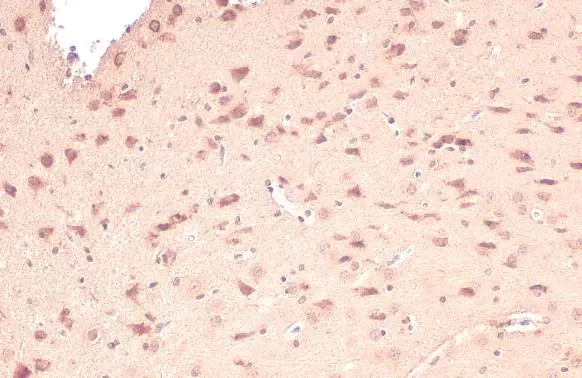
Citrate synthase antibody [N2C3] detects Citrate synthase protein at mitochondria by immunohistochemical analysis. Sample: Paraffin-embedded rat brain. Citrate synthase stained by Citrate synthase antibody [N2C3] (GTX110624) diluted at 1:100. Antigen Retrieval: Citrate buffer, pH 6.0, 15 min
![Citrate synthetase antibody [N2C3] detects Citrate synthetase protein at mitochondria by immunohistochemical analysis. Sample: Paraffin-embedded mouse kidney. Citrate synthetase stained by Citrate synthetase antibody [N2C3] (GTX110624) diluted at 1:500. Antigen Retrieval: Citrate buffer, pH 6.0, 15 min](https://www.genetex.com/upload/website/prouct_img/normal/GTX110624/GTX110624_44664_20220708_IHC-P_M_22071823_202.webp "Citrate synthetase antibody [N2C3] detects Citrate synthetase protein at mitochondria by immunohistochemical analysis. Sample: Paraffin-embedded mouse kidney. Citrate synthetase stained by Citrate synthetase antibody [N2C3] (GTX110624) diluted at 1:500. Antigen Retrieval: Citrate buffer, pH 6.0, 15 min")
![Citrate synthase antibody [N2C3] detects Citrate synthase protein on zebrafish by whole mount immunohistochemical analysis. Sample: 1 day-post-fertilization zebrafish embryo. Citrate synthase antibody [N2C3] (GTX110624) dilution: 1:50.](https://www.genetex.com/upload/website/prouct_img/normal/GTX110624/GTX110624_41773_20151008_IHC-Wm_Z_22111423_697.webp "Citrate synthase antibody [N2C3] detects Citrate synthase protein on zebrafish by whole mount immunohistochemical analysis. Sample: 1 day-post-fertilization zebrafish embryo. Citrate synthase antibody [N2C3] (GTX110624) dilution: 1:50.")
![Various tissue extracts (30 μg) were separated by 10% SDS-PAGE, and the membrane was blotted with Citrate synthase antibody [N2C3] (GTX110624) diluted at 1:500. The HRP-conjugated anti-rabbit IgG antibody (GTX213110-01) was used to detect the primary antibody.](https://www.genetex.com/upload/website/prouct_img/normal/GTX110624/GTX110624_43677_20190913_WB_Z_22111423_856.webp "Various tissue extracts (30 μg) were separated by 10% SDS-PAGE, and the membrane was blotted with Citrate synthase antibody [N2C3] (GTX110624) diluted at 1:500. The HRP-conjugated anti-rabbit IgG antibody (GTX213110-01) was used to detect the primary antibody.")
![Citrate synthase antibody [N2C3] detects Citrate synthase protein on whole mount zebrafish by immunohistochemical analysis. Sample: Paraformaldehyde-fixed 1 day-post-fertilization zebrafish embryo. Green: Citrate synthase stained by Citrate synthase antibody [N2C3] (GTX110624) diluted at 1:100.](https://www.genetex.com/upload/website/prouct_img/normal/GTX110624/GTX110624_43250_20180703_IHC-WM_Z_22111423_838.webp "Citrate synthase antibody [N2C3] detects Citrate synthase protein on whole mount zebrafish by immunohistochemical analysis. Sample: Paraformaldehyde-fixed 1 day-post-fertilization zebrafish embryo. Green: Citrate synthase stained by Citrate synthase antibody [N2C3] (GTX110624) diluted at 1:100.")
 were separated by 10% SDS-PAGE, and the membrane was blotted with Cs antibody (GTX110624) diluted at 1:500. The HRP-conjugated anti-rabbit IgG antibody (GTX213110-01) was used to detect the primary antibody.")
![Citrate synthetase antibody [N2C3] detects Citrate synthetase protein at mitochondria by immunohistochemical analysis. Sample: Paraffin-embedded mouse kidney. Citrate synthetase stained by Citrate synthetase antibody [N2C3] (GTX110624) diluted at 1:500. Antigen Retrieval: Citrate buffer, pH 6.0, 15 min](https://www.genetex.com/upload/website/prouct_img/normal/GTX110624/GTX110624_43901_20200605_IHC-P_M_w_23060500_625.webp "Citrate synthetase antibody [N2C3] detects Citrate synthetase protein at mitochondria by immunohistochemical analysis. Sample: Paraffin-embedded mouse kidney. Citrate synthetase stained by Citrate synthetase antibody [N2C3] (GTX110624) diluted at 1:500. Antigen Retrieval: Citrate buffer, pH 6.0, 15 min")
![Immunoprecipitation of Citrate synthetase protein from 293T whole cell extracts using 5 μg of Citrate synthetase antibody [N2C3] (GTX110624). Western blot analysis was performed using Citrate synthetase antibody [N2C3] (GTX110624). EasyBlot anti-Rabbit IgG (GTX221666-01) was used as a secondary reagent.](https://www.genetex.com/upload/website/prouct_img/normal/GTX110624/GTX110624_41724_20150209_IP_w_23060500_878.webp "Immunoprecipitation of Citrate synthetase protein from 293T whole cell extracts using 5 μg of Citrate synthetase antibody [N2C3] (GTX110624). Western blot analysis was performed using Citrate synthetase antibody [N2C3] (GTX110624). EasyBlot anti-Rabbit IgG (GTX221666-01) was used as a secondary reagent.")
![Citrate synthetase antibody [N2C3] detects Citrate synthetase protein at cytoplasm in mouse muscle by immunohistochemical analysis. Sample: Paraffin-embedded mouse muscle. Citrate synthetase antibody [N2C3] (GTX110624) diluted at 1:500.
Antigen Retrieval: Citrate buffer, pH 6.0, 15 min](https://www.genetex.com/upload/website/prouct_img/normal/GTX110624/GTX110624_41969_20150409_IHC-P_M_2_w_23060500_179.webp "Citrate synthetase antibody [N2C3] detects Citrate synthetase protein at cytoplasm in mouse muscle by immunohistochemical analysis. Sample: Paraffin-embedded mouse muscle. Citrate synthetase antibody [N2C3] (GTX110624) diluted at 1:500.
Antigen Retrieval: Citrate buffer, pH 6.0, 15 min")
![Citrate synthetase antibody [N2C3] detects Citrate synthetase protein at mitochondria by immunohistochemical analysis. Sample: Paraffin-embedded rat heart. Citrate synthetase stained by Citrate synthetase antibody [N2C3] (GTX110624) diluted at 1:500. Antigen Retrieval: Citrate buffer, pH 6.0, 15 min](https://www.genetex.com/upload/website/prouct_img/normal/GTX110624/GTX110624_43901_20200605_IHC-P_R_w_23060500_815.webp "Citrate synthetase antibody [N2C3] detects Citrate synthetase protein at mitochondria by immunohistochemical analysis. Sample: Paraffin-embedded rat heart. Citrate synthetase stained by Citrate synthetase antibody [N2C3] (GTX110624) diluted at 1:500. Antigen Retrieval: Citrate buffer, pH 6.0, 15 min")
Citrate synthase antibody [N2C3] detects Citrate synthase protein at mitochondria by immunohistochemical analysis. Sample: Paraffin-embedded rat brain. Citrate synthase stained by Citrate synthase antibody [N2C3] (GTX110624) diluted at 1:100. Antigen Retrieval: Citrate buffer, pH 6.0, 15 min
Citrate synthase antibody [N2C3]
GTX110624
ApplicationsImmunoFluorescence, ImmunoPrecipitation, Western Blot, ImmunoCytoChemistry, ImmunoHistoChemistry, ImmunoHistoChemistry Frozen, ImmunoHistoChemistry Paraffin
Product group Antibodies
ReactivityAmphibian, Chicken, Human, Monkey, Mouse, Rat, Zebra Fish
TargetCS
Overview
- SupplierGeneTex
- Product NameCitrate synthase antibody [N2C3]
- Delivery Days Customer9
- Application Supplier NoteWB: 1:500-1:3000. ICC/IF: 1:100-1:1000. IHC-P: 1:100-1:1000. IP: 1:100-1:500. *Optimal dilutions/concentrations should be determined by the researcher.Not tested in other applications.
- ApplicationsImmunoFluorescence, ImmunoPrecipitation, Western Blot, ImmunoCytoChemistry, ImmunoHistoChemistry, ImmunoHistoChemistry Frozen, ImmunoHistoChemistry Paraffin
- CertificationResearch Use Only
- ClonalityPolyclonal
- Concentration1.02 mg/ml
- ConjugateUnconjugated
- Gene ID1431
- Target nameCS
- Target descriptioncitrate synthase
- Target synonymscitrate synthase, mitochondrial, citrate (Si)-synthase
- HostRabbit
- IsotypeIgG
- Protein IDO75390
- Protein NameCitrate synthase, mitochondrial
- Scientific DescriptionThe protein encoded by this gene is a Krebs tricarboxylic acid cycle enzyme that catalyzes the synthesis of citrate from oxaloacetate and acetyl coenzyme A. The enzyme is found in nearly all cells capable of oxidative metablism. This protein is nuclear encoded and transported into the mitochondrial matrix, where the mature form is found. [provided by RefSeq]
- ReactivityAmphibian, Chicken, Human, Monkey, Mouse, Rat, Zebra Fish
- Storage Instruction-20°C or -80°C,2°C to 8°C
- UNSPSC41116161
Datasheet
Related products
Product group Antibodies
CS AntibodyCSB-PA006031LA01HU
ApplicationsWestern Blot, ELISA, ImmunoHistoChemistry
ReactivityHuman, Zebra Fish
TargetCS
- SizePrice
Product group Antibodies
ApplicationsDot Blot, ImmunoFluorescence, Western Blot, ELISA
ReactivityPorcine
- SizePrice

Product group Antibodies
Anti-CS AntibodyAMAB91005
ApplicationsWestern Blot, ImmunoCytoChemistry, ImmunoHistoChemistry
ReactivityHuman
TargetCS
- SizePrice

Product group Antibodies
References
ApplicationsFlow Cytometry, ImmunoFluorescence, Western Blot, ImmunoCytoChemistry, ImmunoHistoChemistry
ReactivityHuman, Mouse, Rat
TargetCS
- SizePrice
Product group Antibodies

CS / Citrate Synthase AntibodyLS-C334235
ApplicationsImmunoFluorescence, Western Blot, ImmunoHistoChemistry
ReactivityHuman, Mouse, Rat
TargetCS
- SizePrice
Product group Antibodies
Cs Polyclonal AntibodyCAC07496
ApplicationsWestern Blot, ELISA, ImmunoHistoChemistry
ReactivityZebra Fish
TargetCS
- SizePrice

Product group Antibodies
Citrate synthase Recombinant AntibodyBSM-54190R
ApplicationsFlow Cytometry, ImmunoFluorescence, Western Blot, ImmunoCytoChemistry, ImmunoHistoChemistry, ImmunoHistoChemistry Frozen, ImmunoHistoChemistry Paraffin
ReactivityHuman, Mouse, Rat
TargetCS
- SizePrice
![Citrate synthetase antibody [GT1761] detects Citrate synthetase protein at mitochondria on mouse heart by immunohistochemical analysis. Sample: Paraffin-embedded mouse heart. Citrate synthetase antibody [GT1761] (GTX628143) dilution: 1:200.
Antigen Retrieval: Trilogy? (EDTA based, pH 8.0) buffer, 15min](https://www.genetex.com/upload/website/prouct_img/normal/GTX628143/GTX628143_41113_IHC_M_w_23061202_576.webp)
Product group Antibodies
Citrate synthase antibody [GT1761]GTX628143
ApplicationsImmunoFluorescence, Western Blot, ImmunoCytoChemistry, ImmunoHistoChemistry, ImmunoHistoChemistry Paraffin
ReactivityHuman, Mouse, Rat, Zebra Fish
TargetCS
- SizePrice
![Rat tissue extract (50 μg) was separated by 10% SDS-PAGE, and the membrane was blotted with Citrate synthetase antibody [GT2061] (GTX628144) diluted at 1:1000.](https://www.genetex.com/upload/website/prouct_img/normal/GTX628144/GTX628144_41148_20160114_WB_R_brain_w_23061202_436.webp)
Product group Antibodies
Citrate synthase antibody [GT2061]GTX628144
ApplicationsImmunoFluorescence, Western Blot, ImmunoCytoChemistry, ImmunoHistoChemistry, ImmunoHistoChemistry Paraffin
ReactivityHuman, Mouse, Rat, Zebra Fish
TargetCS
- SizePrice