

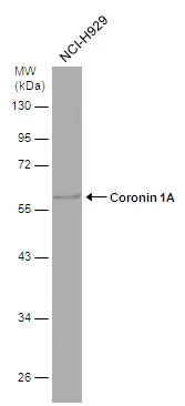
Whole cell extract (30 μg) was separated by 10% SDS-PAGE, and the membrane was blotted with Coronin 1A antibody [N2C2], Internal (GTX113932) diluted at 1:500.
 antibody at 1:100 dilution.
Antigen Retrieval: Trilogy? (EDTA based, pH 8.0) buffer, 15min")
![Mouse tissue extract (50 μg) was separated by 10% SDS-PAGE, and the membrane was blotted with Coronin 1A antibody [N2C2], Internal (GTX113932) diluted at 1:500. The HRP-conjugated anti-rabbit IgG antibody (GTX213110-01) was used to detect the primary antibody, and the signal was developed with Trident ECL plus-Enhanced.](https://www.genetex.com/upload/website/prouct_img/normal/GTX113932/GTX113932_40150_20170713_WB_M_brain_w_23060501_373.webp "Mouse tissue extract (50 μg) was separated by 10% SDS-PAGE, and the membrane was blotted with Coronin 1A antibody [N2C2], Internal (GTX113932) diluted at 1:500. The HRP-conjugated anti-rabbit IgG antibody (GTX213110-01) was used to detect the primary antibody, and the signal was developed with Trident ECL plus-Enhanced.")
Whole cell extract (30 μg) was separated by 10% SDS-PAGE, and the membrane was blotted with Coronin 1A antibody [N2C2], Internal (GTX113932) diluted at 1:500.
Coronin 1A antibody [N2C2], Internal
GTX113932
ApplicationsWestern Blot, ImmunoHistoChemistry, ImmunoHistoChemistry Paraffin
Product group Antibodies
ReactivityHuman, Mouse
TargetCORO1A
Overview
- SupplierGeneTex
- Product NameCoronin 1A antibody [N2C2], Internal
- Delivery Days Customer9
- Application Supplier NoteWB: 1:500-1:3000. IHC-P: 1:100-1:1000. *Optimal dilutions/concentrations should be determined by the researcher.Not tested in other applications.
- ApplicationsWestern Blot, ImmunoHistoChemistry, ImmunoHistoChemistry Paraffin
- CertificationResearch Use Only
- ClonalityPolyclonal
- Concentration1 mg/ml
- ConjugateUnconjugated
- Gene ID11151
- Target nameCORO1A
- Target descriptioncoronin 1A
- Target synonymsCLABP, CLIPINA, HCORO1, IMD8, TACO, p57, coronin-1A, clipin-A, coronin, actin binding protein, 1A, coronin-1, coronin-like protein A, coronin-like protein p57, tryptophan aspartate-containing coat protein
- HostRabbit
- IsotypeIgG
- Protein IDP31146
- Protein NameCoronin-1A
- Scientific DescriptionThis gene encodes a member of the WD repeat protein family. WD repeats are minimally conserved regions of approximately 40 amino acids typically bracketed by gly-his and trp-asp (GH-WD), which may facilitate formation of heterotrimeric or multiprotein complexes. Members of this family are involved in a variety of cellular processes, including cell cycle progression, signal transduction, apoptosis, and gene regulation. Alternative splicing results in multiple transcript variants. A related pseudogene has been defined on chromosome 16. [provided by RefSeq]
- ReactivityHuman, Mouse
- Storage Instruction-20°C or -80°C,2°C to 8°C
- UNSPSC12352203
Datasheet
Related products
Product group Antibodies
Anti-CORO1A Antibody144-09300
ApplicationsImmunoFluorescence, Western Blot
ReactivityHuman, Mouse, Rat
TargetCORO1A
- SizePrice

Product group Antibodies
Anti-Coronin 1a/TACO/CORO1A Antibody Picoband(r)A04245-2-CARRIER-FREE
ApplicationsFlow Cytometry, Western Blot, ELISA, ImmunoHistoChemistry
ReactivityHuman, Mouse, Rat
TargetCORO1A
- SizePrice
![Coronin 1A antibody [C3], C-term detects Coronin 1A protein at cytoplasm in mouse duodenum by immunohistochemical analysis. Sample: Paraffin-embedded mouse duodenum. Coronin 1A antibody [C3], C-term (GTX106424) diluted at 1:500.
Antigen Retrieval: Citrate buffer, pH 6.0, 15 min](https://www.genetex.com/upload/website/prouct_img/normal/GTX106424/GTX106424_39715_20170601_IHC-P_M_2_w_23060120_744.webp)
Product group Antibodies
Coronin 1A antibody [C3], C-termGTX106424
ApplicationsImmunoFluorescence, Western Blot, ImmunoCytoChemistry, ImmunoHistoChemistry, ImmunoHistoChemistry Frozen, ImmunoHistoChemistry Paraffin
ReactivityHuman, Mouse, Rat
TargetCORO1A
- SizePrice
![Coronin 1A antibody [N1N3] detects Coronin 1A protein at cytoplasm in rat intestine by immunohistochemical analysis. Sample: Paraffin-embedded rat intestine. Coronin 1A antibody [N1N3] (GTX113931) diluted at 1:200.](https://www.genetex.com/upload/website/prouct_img/normal/GTX113931/GTX113931_40150_20170601_IHC-P_R_1_w_23060501_269.webp)
Product group Antibodies
Coronin 1A antibody [N1N3]GTX113931
ApplicationsWestern Blot, ImmunoHistoChemistry, ImmunoHistoChemistry Paraffin
ReactivityHuman, Mouse, Rat
TargetCORO1A
- SizePrice
Product group Antibodies
Coronin 1A antibodyGTX85452
ApplicationsImmunoFluorescence, Western Blot, ImmunoCytoChemistry, ImmunoHistoChemistry
ReactivityHuman, Mouse, Rat
TargetCORO1A
- SizePrice
Product group Antibodies
CORO1A Polyclonal AntibodyCAC14636
ApplicationsImmunoFluorescence, Western Blot, ELISA, ImmunoHistoChemistry
ReactivityMouse
TargetCORO1A
- SizePrice
Product group Antibodies
Coronin 1A antibodyGTX14787
ApplicationsWestern Blot, ImmunoHistoChemistry, ImmunoHistoChemistry Paraffin
ReactivityHuman
TargetCORO1A
- SizePrice