

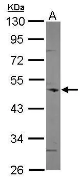
Sample (50 μg of whole cell lysate) A: mouse brain 10% SDS PAGE GTX115727 diluted at 1:500 The HRP-conjugated anti-rabbit IgG antibody (GTX213110-01) was used to detect the primary antibody.
 antibody at 1:500 dilution.")
![Coronin 1C antibody [C1C3] detects Coronin 1C protein at cytoplasm on human lung carcinoma by immunohistochemical analysis. Sample: Paraffin-embedded human lung carcinoma. Coronin 1C antibody [C1C3] (GTX115727) diluted at 1:500.
Antigen Retrieval: Trilogy? (EDTA based, pH 8.0) buffer, 15min](https://www.genetex.com/upload/website/prouct_img/normal/GTX115727/GTX115727_40296_20141205_IHC_w_23060519_539.webp "Coronin 1C antibody [C1C3] detects Coronin 1C protein at cytoplasm on human lung carcinoma by immunohistochemical analysis. Sample: Paraffin-embedded human lung carcinoma. Coronin 1C antibody [C1C3] (GTX115727) diluted at 1:500.
Antigen Retrieval: Trilogy? (EDTA based, pH 8.0) buffer, 15min")
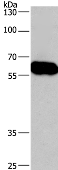
 A: HeLa 10% SDS PAGE GTX115727 diluted at 1:500 The HRP-conjugated anti-rabbit IgG antibody (GTX213110-01) was used to detect the primary antibody.")
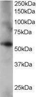
 A: NIH-3T3 10% SDS PAGE GTX115727 diluted at 1:2000 The HRP-conjugated anti-rabbit IgG antibody (GTX213110-01) was used to detect the primary antibody.")
Sample (50 μg of whole cell lysate) A: mouse brain 10% SDS PAGE GTX115727 diluted at 1:500 The HRP-conjugated anti-rabbit IgG antibody (GTX213110-01) was used to detect the primary antibody.
Coronin 1C antibody [C1C3]
GTX115727
ApplicationsImmunoFluorescence, Western Blot, ImmunoCytoChemistry, ImmunoHistoChemistry, ImmunoHistoChemistry Paraffin
Product group Antibodies
ReactivityHuman, Mouse
TargetCORO1C
Overview
- SupplierGeneTex
- Product NameCoronin 1C antibody [C1C3]
- Delivery Days Customer9
- Application Supplier NoteWB: 1:500-1:3000. ICC/IF: 1:100-1:1000. IHC-P: 1:100-1:1000. *Optimal dilutions/concentrations should be determined by the researcher.Not tested in other applications.
- ApplicationsImmunoFluorescence, Western Blot, ImmunoCytoChemistry, ImmunoHistoChemistry, ImmunoHistoChemistry Paraffin
- CertificationResearch Use Only
- ClonalityPolyclonal
- Concentration1 mg/ml
- ConjugateUnconjugated
- Gene ID23603
- Target nameCORO1C
- Target descriptioncoronin 1C
- Target synonymsHCRNN4, coronin-1C, coronin, actin binding protein, 1C, coronin-3
- HostRabbit
- IsotypeIgG
- Protein IDQ9ULV4
- Protein NameCoronin-1C
- Scientific DescriptionThis gene encodes a member of the WD repeat protein family. WD repeats are minimally conserved regions of approximately 40 amino acids typically bracketed by gly-his and trp-asp (GH-WD), which may facilitate formation of heterotrimeric or multiprotein complexes. Members of this family are involved in a variety of cellular processes, including cell cycle progression, signal transduction, apoptosis, and gene regulation. [provided by RefSeq]
- ReactivityHuman, Mouse
- Storage Instruction-20°C or -80°C,2°C to 8°C
- UNSPSC12352203
Datasheet
Related products
Product group Antibodies
Anti-Mouse CORO1C (C-term) Antibody102-23298
ApplicationsWestern Blot
TargetCORO1C
- SizePrice
![Various whole cell extracts (30 μg) were separated by 10% SDS-PAGE, and the membrane was blotted with Coronin 1C antibody [C3], C-term (GTX106469) diluted at 1:1000. The HRP-conjugated anti-rabbit IgG antibody (GTX213110-01) was used to detect the primary antibody, and the signal was developed with Trident ECL plus-Enhanced.](https://www.genetex.com/upload/website/prouct_img/normal/GTX106469/GTX106469_40800_20181026_WB_M_w_23060120_778.webp)
Product group Antibodies
Coronin 1C antibody [C3], C-termGTX106469
ApplicationsImmunoFluorescence, Western Blot, ImmunoCytoChemistry, ImmunoHistoChemistry, ImmunoHistoChemistry Paraffin
ReactivityHuman, Mouse
TargetCORO1C
- SizePrice
Product group Antibodies
CORO1C Polyclonal AntibodyCAC14627
ApplicationsImmunoFluorescence, Western Blot, ELISA
ReactivityMouse
TargetCORO1C
- SizePrice
Product group Antibodies
Coronin 3 Polyclonal AntibodyBS-6799R
ApplicationsImmunoFluorescence, ELISA, ImmunoCytoChemistry, ImmunoHistoChemistry, ImmunoHistoChemistry Frozen, ImmunoHistoChemistry Paraffin
ReactivityBovine, Canine, Equine, Human, Mouse, Porcine, Rabbit, Rat, Sheep
TargetCORO1C
- SizePrice
Product group Antibodies
Anti-CORO1C AntibodyA37211
ApplicationsWestern Blot, ImmunoHistoChemistry
ReactivityHuman
- SizePrice
Product group Antibodies
Coronin 1C antibody, C-termGTX15719
ApplicationsWestern Blot
ReactivityHuman
TargetCORO1C
- SizePrice
Product group Antibodies

ApplicationsWestern Blot, ELISA
ReactivityBovine, Human, Porcine
TargetCORO1C
- SizePrice
Product group Antibodies

CORO1C AntibodyLS-C401542
ApplicationsWestern Blot, ELISA, ImmunoHistoChemistry
ReactivityHuman, Mouse
TargetCORO1C
- SizePrice

Product group Antibodies
Anti-CORO1C AntibodyHPA041737
ApplicationsImmunoHistoChemistry
ReactivityHuman
TargetCORO1C
- SizePrice