

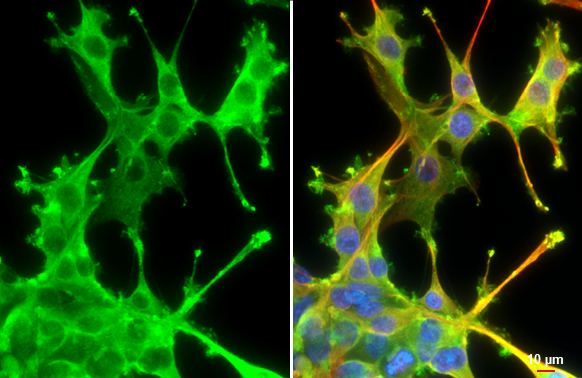
Cortactin antibody [N1], N-term detects Cortactin protein at cytoplasm and cell membrane by immunofluorescent analysis. Sample: U87-MG cells were fixed in 4% paraformaldehyde at RT for 15 min. Green: Cortactin stained by Cortactin antibody [N1], N-term (GTX100253) diluted at 1:500. Red: alpha Tubulin, a cytoskeleton marker, stained by alpha Tubulin antibody [GT114] (GTX628802) diluted at 1:1000. Blue: Fluoroshield with DAPI (GTX30920). Scale bar= 10μm.
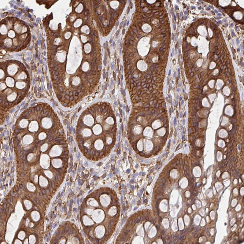
![Cortactin antibody [N1], N-term detects Cortactin protein at cell membrane and cytoplasm by immunohistochemical analysis. Sample: Paraffin-embedded mouse intestine. Cortactin stained by Cortactin antibody [N1], N-term (GTX100253) diluted at 1:500. Antigen Retrieval: Citrate buffer, pH 6.0, 15 min](https://www.genetex.com/upload/website/prouct_img/normal/GTX100253/GTX100253_44482_20211119_IHC-P_M_w_23060100_565.webp "Cortactin antibody [N1], N-term detects Cortactin protein at cell membrane and cytoplasm by immunohistochemical analysis. Sample: Paraffin-embedded mouse intestine. Cortactin stained by Cortactin antibody [N1], N-term (GTX100253) diluted at 1:500. Antigen Retrieval: Citrate buffer, pH 6.0, 15 min")
 A: PC-12 7.5% SDS PAGE GTX100253 diluted at 1:3000")
 antibody at 1:250 dilution.
Antigen Retrieval: Trilogy? (EDTA based, pH 8.0) buffer, 15min")
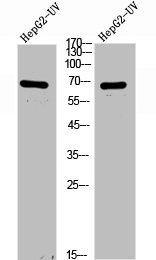
![Wild-type (WT) and Cortactin knockout (KO) HeLa cell extracts (30 μg) were separated by 7.5% SDS-PAGE, and the membrane was blotted with Cortactin antibody [N1], N-term (GTX100253) diluted at 1:1000. The HRP-conjugated anti-rabbit IgG antibody (GTX213110-01) was used to detect the primary antibody.](https://www.genetex.com/upload/website/prouct_img/normal/GTX100253/GTX100253_39568_20170309_WB_KO_watermark_w_23060100_838.webp "Wild-type (WT) and Cortactin knockout (KO) HeLa cell extracts (30 μg) were separated by 7.5% SDS-PAGE, and the membrane was blotted with Cortactin antibody [N1], N-term (GTX100253) diluted at 1:1000. The HRP-conjugated anti-rabbit IgG antibody (GTX213110-01) was used to detect the primary antibody.")
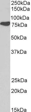
 A: NIH-3T3 B: JC 7.5% SDS PAGE GTX100253 diluted at 1:1000")
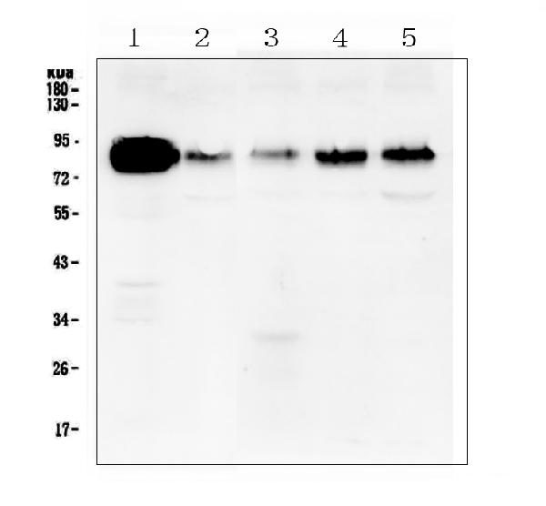
![Various whole cell extracts (30 μg) were separated by 7.5% SDS-PAGE, and the membrane was blotted with Cortactin antibody [N1], N-term (GTX100253) diluted at 1:1000. The HRP-conjugated anti-rabbit IgG antibody (GTX213110-01) was used to detect the primary antibody.](https://www.genetex.com/upload/website/prouct_img/normal/GTX100253/GTX100253_44482_20211015_WB_w_23060100_870.webp "Various whole cell extracts (30 μg) were separated by 7.5% SDS-PAGE, and the membrane was blotted with Cortactin antibody [N1], N-term (GTX100253) diluted at 1:1000. The HRP-conjugated anti-rabbit IgG antibody (GTX213110-01) was used to detect the primary antibody.")
![Various tissue extracts (50 μg) were separated by 7.5% SDS-PAGE, and the membrane was blotted with Cortactin antibody [N1], N-term (GTX100253) diluted at 1:1000. The HRP-conjugated anti-rabbit IgG antibody (GTX213110-01) was used to detect the primary antibody.](https://www.genetex.com/upload/website/prouct_img/normal/GTX100253/GTX100253_39568_20170518_WB_M_R_w_23060100_747.webp "Various tissue extracts (50 μg) were separated by 7.5% SDS-PAGE, and the membrane was blotted with Cortactin antibody [N1], N-term (GTX100253) diluted at 1:1000. The HRP-conjugated anti-rabbit IgG antibody (GTX213110-01) was used to detect the primary antibody.")
Cortactin antibody [N1], N-term detects Cortactin protein at cytoplasm and cell membrane by immunofluorescent analysis. Sample: U87-MG cells were fixed in 4% paraformaldehyde at RT for 15 min. Green: Cortactin stained by Cortactin antibody [N1], N-term (GTX100253) diluted at 1:500. Red: alpha Tubulin, a cytoskeleton marker, stained by alpha Tubulin antibody [GT114] (GTX628802) diluted at 1:1000. Blue: Fluoroshield with DAPI (GTX30920). Scale bar= 10μm.
Cortactin antibody [N1], N-term
GTX100253
ApplicationsImmunoFluorescence, Western Blot, ImmunoCytoChemistry, ImmunoHistoChemistry, ImmunoHistoChemistry Paraffin
Product group Antibodies
ReactivityHuman, Mouse, Rat
TargetCTTN
Overview
- SupplierGeneTex
- Product NameCortactin antibody [N1], N-term
- Delivery Days Customer9
- Application Supplier NoteWB: 1:500-1:3000. ICC/IF: 1:100-1:1000. IHC-P: 1:100-1:1000. *Optimal dilutions/concentrations should be determined by the researcher.Not tested in other applications.
- ApplicationsImmunoFluorescence, Western Blot, ImmunoCytoChemistry, ImmunoHistoChemistry, ImmunoHistoChemistry Paraffin
- CertificationResearch Use Only
- ClonalityPolyclonal
- Concentration1 mg/ml
- ConjugateUnconjugated
- Gene ID2017
- Target nameCTTN
- Target descriptioncortactin
- Target synonymsEMS1, src substrate cortactin, amplaxin, ems1 sequence (mammary tumor and squamous cell carcinoma-associated (p80/85 src substrate), epididymis secretory sperm binding protein, oncogene EMS1
- HostRabbit
- IsotypeIgG
- Protein IDQ14247
- Protein NameSrc substrate cortactin
- Scientific DescriptionThis gene is overexpressed in breast cancer and squamous cell carcinomas of the head and neck. The encoded protein is localized in the cytoplasm and in areas of the cell-substratum contacts. This gene has two roles: (1) regulating the interactions between components of adherens-type junctions and (2) organizing the cytoskeleton and cell adhesion structures of epithelia and carcinoma cells. During apoptosis, the encoded protein is degraded in a caspase-dependent manner. The aberrant regulation of this gene contributes to tumor cell invasion and metastasis. Two splice variants that encode different isoforms have been identified for this gene. [provided by RefSeq]
- ReactivityHuman, Mouse, Rat
- Storage Instruction-20°C or -80°C,2°C to 8°C
- UNSPSC41116161
Datasheet
Related products
Product group Antibodies
Anti-Cortactin AntibodyA96003
ApplicationsWestern Blot, ELISA
ReactivityHuman, Mouse, Rat
- SizePrice
Product group Antibodies
Anti-Cortactin/CTTN Antibody Picoband(r)A01253-1-CARRIER-FREE
ApplicationsFlow Cytometry, ImmunoFluorescence, Western Blot, ELISA, ImmunoCytoChemistry, ImmunoHistoChemistry
ReactivityHuman, Mouse, Rat
TargetCTTN
- SizePrice
Product group Antibodies
Anti-CTTN Antibody144-60898
ApplicationsWestern Blot
ReactivityHuman, Mouse, Rat
TargetCTTN
- SizePrice
Product group Antibodies

CTTN / Cortactin AntibodyLS-C750030
ApplicationsWestern Blot
ReactivityHuman, Mouse, Rat
TargetCTTN
- SizePrice
Product group Antibodies
Cortactin Recombinant AntibodyBSM-61251R
ApplicationsFlow Cytometry, ImmunoFluorescence, ImmunoPrecipitation, Western Blot, ImmunoCytoChemistry, ImmunoHistoChemistry, ImmunoHistoChemistry Frozen, ImmunoHistoChemistry Paraffin
TargetCTTN
- SizePrice
Product group Antibodies
CTTN AntibodyCSB-PA006097
ApplicationsWestern Blot, ELISA
ReactivityHuman, Mouse, Rat
TargetCTTN
- SizePrice
Product group Antibodies

Goat anti-Cortactin / EMS1EB05253
ApplicationsWestern Blot, ELISA
ReactivityBovine, Canine, Human, Mouse, Rat
TargetCTTN
- SizePrice
Product group Antibodies
Cortactin antibody, N-termGTX21374
ApplicationsWestern Blot
ReactivityHuman, Mouse, Rat
TargetCTTN
- SizePrice
Product group Antibodies
Anti-CTTN AntibodyHPA057242
ApplicationsWestern Blot, ImmunoCytoChemistry, ImmunoHistoChemistry
ReactivityHuman
TargetCTTN
- SizePrice