

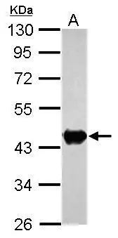
Sample (30 μg of whole cell lysate) A: zebrafish eye 10% SDS PAGE GTX101759 diluted at 1:1000
![Immunohistochemical analysis of paraffin-embedded zebrafish tissue, using Creatine kinase (brain) antibody [N1C1] (GTX101759) at 1:300 dilution.](https://www.genetex.com/upload/website/prouct_img/normal/GTX101759/GTX101759_39577_IHC_Z_22111423_340.webp "Immunohistochemical analysis of paraffin-embedded zebrafish tissue, using Creatine kinase (brain) antibody [N1C1] (GTX101759) at 1:300 dilution.")
![Creatine kinase (brain) antibody [N1C1] detects Creatine kinase (brain) protein at cytoplasm by immunofluorescent analysis. Sample: 293T cells were fixed in 4% paraformaldehyde at RT for 15 min. Green: Creatine kinase (brain) stained by Creatine kinase (brain) antibody [N1C1] (GTX101759) diluted at 1:1000. Blue: Fluoroshield with DAPI (GTX30920).](https://www.genetex.com/upload/website/prouct_img/normal/GTX101759/GTX101759_43755_20200812_ICC_IF_w_23060100_341.webp "Creatine kinase (brain) antibody [N1C1] detects Creatine kinase (brain) protein at cytoplasm by immunofluorescent analysis. Sample: 293T cells were fixed in 4% paraformaldehyde at RT for 15 min. Green: Creatine kinase (brain) stained by Creatine kinase (brain) antibody [N1C1] (GTX101759) diluted at 1:1000. Blue: Fluoroshield with DAPI (GTX30920).")
![Creatine kinase (brain) antibody [N1C1] detects Creatine kinase (brain) protein at cytoplasm by immunohistochemical analysis. Sample: Paraffin-embedded mouse intestine. Creatine kinase (brain) stained by Creatine kinase (brain) antibody [N1C1] (GTX101759) diluted at 1:500. Antigen Retrieval: Citrate buffer, pH 6.0, 15 min](https://www.genetex.com/upload/website/prouct_img/normal/GTX101759/GTX101759_43754_20200731_IHC-P_M_w_23060100_727.webp "Creatine kinase (brain) antibody [N1C1] detects Creatine kinase (brain) protein at cytoplasm by immunohistochemical analysis. Sample: Paraffin-embedded mouse intestine. Creatine kinase (brain) stained by Creatine kinase (brain) antibody [N1C1] (GTX101759) diluted at 1:500. Antigen Retrieval: Citrate buffer, pH 6.0, 15 min")
![Creatine kinase (brain) antibody [N1C1] detects Creatine kinase (brain) protein at cytoplasm and nucleus by immunofluorescent analysis. Sample: HeLa cells were fixed in 4% paraformaldehyde at RT for 15 min. Green: Creatine kinase (brain) protein stained by Creatine kinase (brain) antibody [N1C1] (GTX101759) diluted at 1:500. Red: alpha Tubulin, a cytoskeleton marker, stained by alpha Tubulin antibody [B-5-1-2] (GTX11304) diluted at 1:10000. Blue: Hoechst 33342 staining.](https://www.genetex.com/upload/website/prouct_img/normal/GTX101759/GTX101759_39577_20150410_IFA_w_23060100_675.webp "Creatine kinase (brain) antibody [N1C1] detects Creatine kinase (brain) protein at cytoplasm and nucleus by immunofluorescent analysis. Sample: HeLa cells were fixed in 4% paraformaldehyde at RT for 15 min. Green: Creatine kinase (brain) protein stained by Creatine kinase (brain) antibody [N1C1] (GTX101759) diluted at 1:500. Red: alpha Tubulin, a cytoskeleton marker, stained by alpha Tubulin antibody [B-5-1-2] (GTX11304) diluted at 1:10000. Blue: Hoechst 33342 staining.")
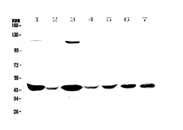
![Various tissue extracts (50 μg) were separated by 10% SDS-PAGE, and the membrane was blotted with Creatine kinase (brain) antibody [N1C1] (GTX101759) diluted at 1:1000. The HRP-conjugated anti-rabbit IgG antibody (GTX213110-01) was used to detect the primary antibody.](https://www.genetex.com/upload/website/prouct_img/normal/GTX101759/GTX101759_43586_20190517_WB_M_R_w_23060100_120.webp "Various tissue extracts (50 μg) were separated by 10% SDS-PAGE, and the membrane was blotted with Creatine kinase (brain) antibody [N1C1] (GTX101759) diluted at 1:1000. The HRP-conjugated anti-rabbit IgG antibody (GTX213110-01) was used to detect the primary antibody.")
 antibody(GTX101759) antibody at 1:500 dilution.
Antigen Retrieval: Trilogy? (EDTA based, pH 8.0) buffer, 15min")
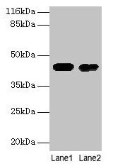
![Various whole cell extracts (30 μg) were separated by 10% SDS-PAGE, and the membrane was blotted with Creatine kinase (brain) antibody [N1C1] (GTX101759) diluted at 1:1000. The HRP-conjugated anti-rabbit IgG antibody (GTX213110-01) was used to detect the primary antibody.](https://www.genetex.com/upload/website/prouct_img/normal/GTX101759/GTX101759_43621_20191101_WB_24112622_585.webp "Various whole cell extracts (30 μg) were separated by 10% SDS-PAGE, and the membrane was blotted with Creatine kinase (brain) antibody [N1C1] (GTX101759) diluted at 1:1000. The HRP-conjugated anti-rabbit IgG antibody (GTX213110-01) was used to detect the primary antibody.")
Sample (30 μg of whole cell lysate) A: zebrafish eye 10% SDS PAGE GTX101759 diluted at 1:1000
Creatine kinase (brain) antibody [N1C1]
GTX101759
ApplicationsImmunoFluorescence, Western Blot, ImmunoCytoChemistry, ImmunoHistoChemistry, ImmunoHistoChemistry Paraffin
Product group Antibodies
ReactivityHuman, Mouse, Rat, Zebra Fish
TargetCKB
Overview
- SupplierGeneTex
- Product NameCreatine kinase (brain) antibody [N1C1]
- Delivery Days Customer9
- Application Supplier NoteWB: 1:500-1:3000. ICC/IF: 1:100-1:1000. IHC-P: 1:100-1:1000. *Optimal dilutions/concentrations should be determined by the researcher.Not tested in other applications.
- ApplicationsImmunoFluorescence, Western Blot, ImmunoCytoChemistry, ImmunoHistoChemistry, ImmunoHistoChemistry Paraffin
- CertificationResearch Use Only
- ClonalityPolyclonal
- Concentration1.91 mg/ml
- ConjugateUnconjugated
- Gene ID1152
- Target nameCKB
- Target descriptioncreatine kinase B
- Target synonymsB-CK, BCK, CKBB, CPK-B, HEL-211, HEL-S-29, creatine kinase B-type, brain creatine kinase, creatine kinase B chain, creatine kinase brain, creatine kinase brain-type, creatine phosphokinase B-type, epididymis luminal protein 211, epididymis secretory protein Li 29
- HostRabbit
- IsotypeIgG
- Protein IDP12277
- Protein NameCreatine kinase B-type
- Scientific DescriptionThe protein encoded by this gene is a cytoplasmic enzyme involved in energy homeostasis. The encoded protein reversibly catalyzes the transfer of phosphate between ATP and various phosphogens such as creatine phosphate. It acts as a homodimer in brain as well as in other tissues, and as a heterodimer with a similar muscle isozyme in heart. The encoded protein is a member of the ATP:guanido phosphotransferase protein family. A pseudogene of this gene has been characterized. [provided by RefSeq]
- ReactivityHuman, Mouse, Rat, Zebra Fish
- Storage Instruction-20°C or -80°C,2°C to 8°C
- UNSPSC41116161
Datasheet
Related products
Product group Antibodies
Anti-CKB Antibody130-10062
ApplicationsWestern Blot, ELISA
ReactivityHuman
TargetCKB
- SizePrice

Product group Antibodies

ApplicationsELISA
ReactivityHuman
TargetCKB
- SizePrice
Product group Antibodies
Anti-Creatine kinase B type/CKB Antibody Picoband(r)A01695-1-CARRIER-FREE
ApplicationsWestern Blot, ELISA, ImmunoHistoChemistry
ReactivityHuman, Mouse, Rat
TargetCKB
- SizePrice
Product group Antibodies
ApplicationsFlow Cytometry, ImmunoFluorescence, Western Blot
ReactivityHuman, Mouse, Rat
TargetCKB
- SizePrice
Product group Antibodies

ApplicationsWestern Blot, ELISA
ReactivityBovine, Canine, Human, Mouse, Porcine, Rat
TargetCKB
- SizePrice
Product group Antibodies
CKB Polyclonal AntibodyCAC13835
ApplicationsImmunoFluorescence, Western Blot, ELISA, ImmunoHistoChemistry
ReactivityMouse
TargetCKB
- SizePrice
Product group Antibodies
CKB AntibodyCSB-PA01495A0RB
ApplicationsImmunoFluorescence, Western Blot, ELISA, ImmunoHistoChemistry
ReactivityHuman, Mouse
TargetCKB
- SizePrice
![Immunohistochemical analysis of paraffin-embedded zebrafish tissue, using Creatine kinase (brain) antibody [N3C3] (GTX101760) at 1:300 dilution.](https://www.genetex.com/upload/website/prouct_img/normal/GTX101760/GTX101760_39827_IHC_Z_22111422_980.webp)
Product group Antibodies
ApplicationsImmunoFluorescence, Western Blot, ImmunoCytoChemistry, ImmunoHistoChemistry, ImmunoHistoChemistry Paraffin
ReactivityHuman, Mouse, Rat, Zebra Fish
TargetCKB
- SizePrice
Product group Antibodies
ApplicationsWestern Blot
ReactivityHuman
TargetCKB
- SizePrice