

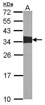
Sample (30 ug of whole cell lysate) A:NIH-3T3 12% SDS PAGE GTX100901 diluted at 1:1000
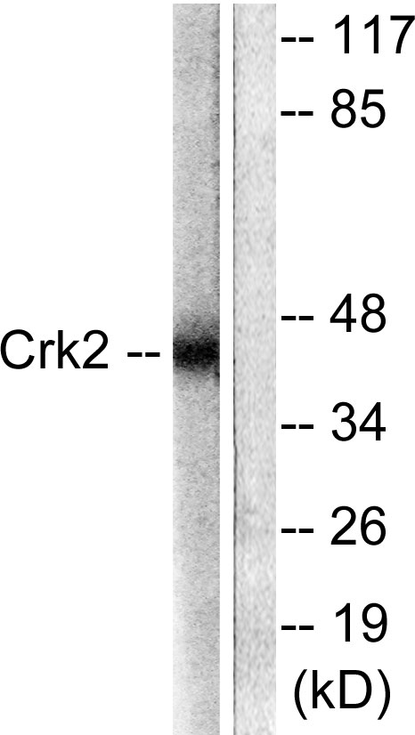
 antibody at 1:200 dilution.")
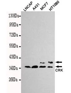
![Various whole cell extracts (30 μg) were separated by 12% SDS-PAGE, and the membranes were blotted with CRK antibody [N2C3] (GTX100901) diluted at 1:1000 and competitor's antibody (SC-289) diluted by 1:200.](https://www.genetex.com/upload/website/prouct_img/normal/GTX100901/GTX100901_39596_20161124_WB_competitor_watermark_w_23060100_456.webp "Various whole cell extracts (30 μg) were separated by 12% SDS-PAGE, and the membranes were blotted with CRK antibody [N2C3] (GTX100901) diluted at 1:1000 and competitor's antibody (SC-289) diluted by 1:200.")
Sample (30 ug of whole cell lysate) A:NIH-3T3 12% SDS PAGE GTX100901 diluted at 1:1000
CRK antibody [N2C3]
GTX100901
ApplicationsImmunoFluorescence, Western Blot, ImmunoCytoChemistry
Product group Antibodies
ReactivityHuman, Mouse
TargetCRK
Overview
- SupplierGeneTex
- Product NameCRK antibody [N2C3]
- Delivery Days Customer9
- Application Supplier NoteWB: 1:500-1:3000. ICC/IF: 1:100-1:1000. *Optimal dilutions/concentrations should be determined by the researcher.Not tested in other applications.
- ApplicationsImmunoFluorescence, Western Blot, ImmunoCytoChemistry
- CertificationResearch Use Only
- ClonalityPolyclonal
- Concentration1 mg/ml
- ConjugateUnconjugated
- Gene ID1398
- Target nameCRK
- Target descriptionCRK proto-oncogene, adaptor protein
- Target synonymsCRKII, p38, adapter molecule crk, proto-oncogene c-Crk, v-crk avian sarcoma virus CT10 oncogene homolog, v-crk sarcoma virus CT10 oncogene-like protein
- HostRabbit
- IsotypeIgG
- Protein IDP46108
- Protein NameAdapter molecule crk
- Scientific DescriptionThis gene encodes a member of an adapter protein family that binds to several tyrosine-phosphorylated proteins. The product of this gene has several SH2 and SH3 domains (src-homology domains) and is involved in several signaling pathways, recruiting cytoplasmic proteins in the vicinity of tyrosine kinase through SH2-phosphotyrosine interaction. The N-terminal SH2 domain of this protein functions as a positive regulator of transformation whereas the C-terminal SH3 domain functions as a negative regulator of transformation. Two alternative transcripts encoding different isoforms with distinct biological activity have been described. [provided by RefSeq]
- ReactivityHuman, Mouse
- Storage Instruction-20°C or -80°C,2°C to 8°C
- UNSPSC41116161
Datasheet
Related products
Product group Antibodies
Anti-CrkII AntibodyA95240
ApplicationsImmunoFluorescence, Western Blot, ELISA, ImmunoHistoChemistry
ReactivityHuman, Mouse, Rat
- SizePrice
Product group Antibodies
Anti-CRK Antibody144-01604
ApplicationsWestern Blot, ImmunoHistoChemistry
ReactivityHuman, Mouse
TargetCRK
- SizePrice
Product group Antibodies
ApplicationsWestern Blot
ReactivityHuman
TargetCRK
- SizePrice
Product group Antibodies
CRK AntibodyCSB-PA001789
ApplicationsImmunoFluorescence, Western Blot, ELISA, ImmunoHistoChemistry
ReactivityHuman, Monkey, Mouse, Rat
TargetCRK
- SizePrice
Product group Antibodies
CRK Polyclonal AntibodyCAC14956
ApplicationsImmunoFluorescence, Western Blot, ELISA
ReactivityRat
TargetCRK
- SizePrice
Product group Antibodies

CRK AntibodyLS-C401551
ApplicationsELISA, ImmunoHistoChemistry
ReactivityHuman, Mouse, Rat
TargetCRK
- SizePrice
Product group Antibodies
CRK antibodyGTX78919
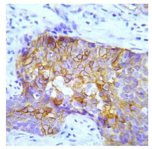
ApplicationsImmunoHistoChemistry, ImmunoHistoChemistry Paraffin
ReactivityHuman
TargetCRK
- SizePrice
Product group Antibodies
CRK (phospho Tyr221) antibodyGTX79097
ApplicationsImmunoHistoChemistry, ImmunoHistoChemistry Paraffin
ReactivityHuman
TargetCRK
- SizePrice
Product group Antibodies
Anti-CRK AntibodyHPA068087
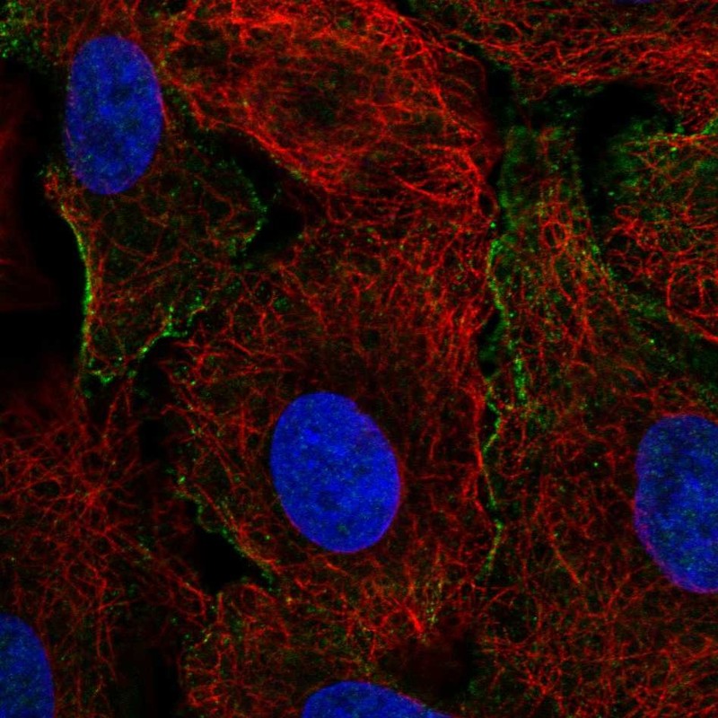
ApplicationsImmunoCytoChemistry
ReactivityHuman
TargetCRK
- SizePrice
Product group Antibodies
CRK antibody [3H7-E5-H8]GTX49212
ApplicationsWestern Blot
ReactivityHuman
TargetCRK
- SizePrice