

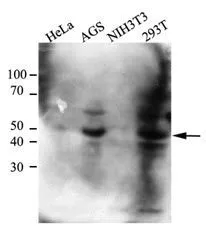
WB analysis of the indicated lysates (40ug per lane) using Cystatin B antibody [2F1] at a dilution of 1:500.
WB analysis of the indicated lysates (40ug per lane) using Cystatin B antibody [2F1] at a dilution of 1:500.
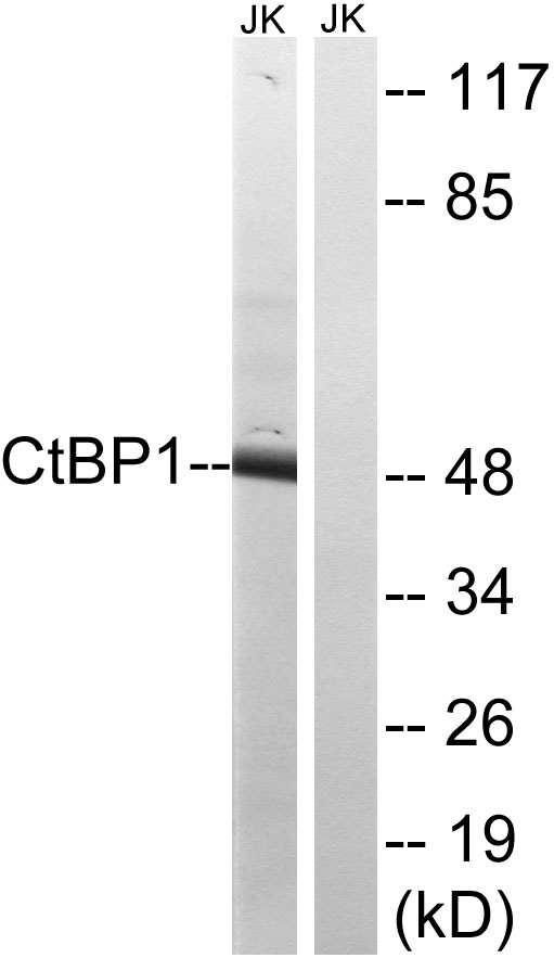
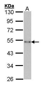
CtBP1 antibody [AT4D6]
GTX53708
ApplicationsWestern Blot, ELISA
Product group Antibodies
ReactivityHuman
TargetCTBP1
Overview
- SupplierGeneTex
- Product NameCtBP1 antibody [AT4D6]
- Delivery Days Customer9
- Application Supplier NoteThe antibody has been tested by ELISA and Western blot analysis to assure specificity and reactivity. Since application varies, however, each investigation should be titrated by the reagent to obtain optimal results. Recommended dilution range for Western blot analysis is 1:500. Recommended starting dilution is 1:500
- ApplicationsWestern Blot, ELISA
- CertificationResearch Use Only
- ClonalityMonoclonal
- Clone IDAT4D6
- Concentration1 mg/ml
- ConjugateUnconjugated
- Gene ID1487
- Target nameCTBP1
- Target descriptionC-terminal binding protein 1
- Target synonymsBARS, HADDTS, C-terminal-binding protein 1, brefeldin A-ribosylated substrate
- HostMouse
- IsotypeIgG2b
- Protein IDQ13363
- Protein NameC-terminal-binding protein 1
- Scientific DescriptionThis gene encodes a protein that binds to the C-terminus of adenovirus E1A proteins. This phosphoprotein is a transcriptional repressor and may play a role during cellular proliferation. This protein and the product of a second closely related gene, CTBP2, can dimerize. Both proteins can also interact with a polycomb group protein complex which participates in regulation of gene expression during development. Alternative splicing of transcripts from this gene results in multiple transcript variants. [provided by RefSeq, Jul 2008]
- ReactivityHuman
- Storage Instruction-20°C or -80°C,2°C to 8°C
- UNSPSC41116161
Datasheet
Related products
Product group Antibodies
Anti-CtBP1 AntibodyA95895
ApplicationsWestern Blot, ELISA, ImmunoHistoChemistry
ReactivityHuman, Mouse, Rat
- SizePrice
Product group Antibodies

CtBP1 (Phospho-Ser422) AntibodyABX012683
ApplicationsWestern Blot, ELISA, ImmunoHistoChemistry
- SizePrice
Product group Antibodies
Anti-CTBP1 Antibody144-01707
ApplicationsWestern Blot, ImmunoHistoChemistry
ReactivityHuman, Mouse
TargetCTBP1
- SizePrice
Product group Antibodies

CTBP1 / CTBP AntibodyLS-C765865
ApplicationsELISA, ImmunoHistoChemistry
ReactivityHuman, Mouse, Rat
TargetCTBP1
- SizePrice
Product group Antibodies
CTBP1 Recombinant Antibody, AbBy Fluor-405 ConjugatedBSM-61953R-BF405
ApplicationsImmunoFluorescence, Western Blot
ReactivityHuman, Mouse, Rat
TargetCTBP1
- SizePrice

Product group Antibodies
Phospho-CTBP1 (S422) AntibodyCSB-PA007410
ApplicationsWestern Blot, ELISA, ImmunoHistoChemistry
ReactivityHuman, Mouse, Rat
TargetCTBP1
- SizePrice
Product group Antibodies
ApplicationsImmunoPrecipitation, Western Blot, ImmunoCytoChemistry, ImmunoHistoChemistry
ReactivityMouse, Rat
TargetCTBP1
- SizePrice
Product group Antibodies
CtBP1 antibodyGTX14411
ApplicationsImmunoFluorescence, ImmunoPrecipitation, Western Blot, ImmunoCytoChemistry
ReactivityHuman, Mouse
TargetCTBP1
- SizePrice
Product group Antibodies
CtBP1 antibody [N3C3]GTX101775
ApplicationsImmunoFluorescence, Western Blot, ImmunoCytoChemistry
ReactivityHuman
TargetCTBP1
- SizePrice
Product group Antibodies
CtBP1 antibody [N2C2], InternalGTX111229
ApplicationsImmunoFluorescence, Western Blot, ImmunoCytoChemistry, ImmunoHistoChemistry
ReactivityHuman, Mouse, Zebra Fish
TargetCTBP1
- SizePrice