

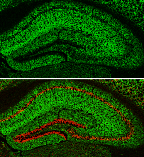
MAP2 antibody [HM-2] detects MAP2 protein expression by immunohistochemical analysis. Sample: Frozen-sectioned adult mouse hippocampus. Green: MAP2 protein stained by MAP2 antibody [HM-2] (GTX11267) diluted at 1:250. Red: NeuN, stained by NeuN antibody (GTX132974) diluted at 1:500.
![MAP2 antibody [HM-2] detects MAP2 protein at cytoplasm in mouse brain by immunohistochemical analysis. Sample: Paraffin-embedded mouse brain. MAP2 antibody [HM-2] (GTX11267) diluted at 1:500.](https://www.genetex.com/upload/website/prouct_img/normal/GTX11267/GTX11267_20161129_IHC-P_M_w_23060500_252.webp "MAP2 antibody [HM-2] detects MAP2 protein at cytoplasm in mouse brain by immunohistochemical analysis. Sample: Paraffin-embedded mouse brain. MAP2 antibody [HM-2] (GTX11267) diluted at 1:500.")
![WB analysis of rat brain extract using GTX11267 MAP2 antibody [HM-2]at 1 μg/mL.](https://www.genetex.com/upload/website/prouct_img/normal/GTX11267/GTX11267_20170605_WB_w_23060500_894.webp "WB analysis of rat brain extract using GTX11267 MAP2 antibody [HM-2]at 1 μg/mL.")
![MAP2 antibody [HM-2] detects MAP2 protein at by immunofluorescent analysis. Sample: Rat E18 primary cortical neuron, DIV 8. Cells were fixed in 4% paraformaldehyde at RT for 15 min. Green: MAP2 protein stained by MAP2 antibody [HM-2] (GTX11267) diluted at 1:250. Red: NeuN, stained by NeuN antibody (GTX132974) diluted at 1:250.](https://www.genetex.com/upload/website/prouct_img/normal/GTX11267/GTX11267__20161024_IFA_R_w_23060500_471.webp "MAP2 antibody [HM-2] detects MAP2 protein at by immunofluorescent analysis. Sample: Rat E18 primary cortical neuron, DIV 8. Cells were fixed in 4% paraformaldehyde at RT for 15 min. Green: MAP2 protein stained by MAP2 antibody [HM-2] (GTX11267) diluted at 1:250. Red: NeuN, stained by NeuN antibody (GTX132974) diluted at 1:250.")
![MAP2 antibody [HM-2] detects MAP2 protein at cytoplasm in rat brain by immunohistochemical analysis. Sample: Paraffin-embedded rat brain. MAP2 antibody [HM-2] (GTX11267) diluted at 1:500.](https://www.genetex.com/upload/website/prouct_img/normal/GTX11267/GTX11267_20161129_IHC-P_R_w_23060500_671.webp "MAP2 antibody [HM-2] detects MAP2 protein at cytoplasm in rat brain by immunohistochemical analysis. Sample: Paraffin-embedded rat brain. MAP2 antibody [HM-2] (GTX11267) diluted at 1:500.")
![ICC/IF analysis of B35 cells using GTX11267 MAP2 antibody [HM-2] at 2 μg/mL(red) with DAPI(blue). Cells were fixed and permeabilized with methanol followed by methanol:acetone.](https://www.genetex.com/upload/website/prouct_img/normal/GTX11267/GTX11267_20170605_ICCIF_w_23060500_779.webp "ICC/IF analysis of B35 cells using GTX11267 MAP2 antibody [HM-2] at 2 μg/mL(red) with DAPI(blue). Cells were fixed and permeabilized with methanol followed by methanol:acetone.")
![MAP2 antibody [HM-2] detects MAP2 protein expression by immunohistochemical analysis. Sample: Frozen-sectioned adult mouse cerebellum. Green: MAP2 protein stained by MAP2 antibody [HM-2] (GTX11267) diluted at 1:250. Blue: Fluoroshield with DAPI (GTX30920).](https://www.genetex.com/upload/website/prouct_img/normal/GTX11267/GTX11267_20170531_IHC-Fr_M_w_23060500_427.webp "MAP2 antibody [HM-2] detects MAP2 protein expression by immunohistochemical analysis. Sample: Frozen-sectioned adult mouse cerebellum. Green: MAP2 protein stained by MAP2 antibody [HM-2] (GTX11267) diluted at 1:250. Blue: Fluoroshield with DAPI (GTX30920).")
![Various whole cell extracts (30 μg) were separated by 7.5% SDS-PAGE, and the membrane was blotted with MAP2 antibody [HM-2] (GTX11267) diluted at 1:2000. The signal was developed with Trident ECL plus-Enhanced.](https://www.genetex.com/upload/website/prouct_img/normal/GTX11267/GTX11267_821603282_20161208_WB_R_w_23060500_762.webp "Various whole cell extracts (30 μg) were separated by 7.5% SDS-PAGE, and the membrane was blotted with MAP2 antibody [HM-2] (GTX11267) diluted at 1:2000. The signal was developed with Trident ECL plus-Enhanced.")
MAP2 antibody [HM-2] detects MAP2 protein expression by immunohistochemical analysis. Sample: Frozen-sectioned adult mouse hippocampus. Green: MAP2 protein stained by MAP2 antibody [HM-2] (GTX11267) diluted at 1:250. Red: NeuN, stained by NeuN antibody (GTX132974) diluted at 1:500.
MAP2 antibody [HM-2]
GTX11267
ApplicationsImmunoFluorescence, Western Blot, ImmunoCytoChemistry, ImmunoHistoChemistry, ImmunoHistoChemistry Frozen, ImmunoHistoChemistry Paraffin
Product group Antibodies
ReactivityAvian, Bovine, Chicken, Human, Mouse, Rat
TargetMap2
Overview
- SupplierGeneTex
- Product NameMAP2 antibody [HM-2]
- Delivery Days Customer9
- Application Supplier NoteWB: 1-2 microg/ml. IHC-P: 1:100-1:1000. IHC-Fr: 1:100-1:1000. *Optimal dilutions/concentrations should be determined by the researcher.Not tested in other applications.
- ApplicationsImmunoFluorescence, Western Blot, ImmunoCytoChemistry, ImmunoHistoChemistry, ImmunoHistoChemistry Frozen, ImmunoHistoChemistry Paraffin
- CertificationResearch Use Only
- ClonalityMonoclonal
- Clone IDHM-2
- Concentration2 mg/ml
- ConjugateUnconjugated
- Gene ID25595
- Target nameMap2
- Target descriptionmicrotubule-associated protein 2
- Target synonymsMAP2R, Mtap2, microtubule-associated protein 2, microtubule-associated protein 2C
- HostMouse
- IsotypeIgG1
- Protein IDP15146
- Protein NameMicrotubule-associated protein 2
- Scientific DescriptionMAP2 is the major microtubule associated protein of brain tissue. There are three forms of MAP2; two are similarily sized with apparent molecular weights of 280 kDa (MAP2a and MAP2b) and the third with a lower molecular weight of 70 kDa (MAP2c). In the newborn rat brain, MAP2b and MAP2c are present, while MAP2a is absent. Between postnatal days 10 and 20, MAP2a appears. At the same time, the level of MAP2c drops by 10-fold. This change happens during the period when dendrite growth is completed and when neurons have reached their mature morphology. MAP2 is degraded by a Cathepsin D-like protease in the brain of aged rats. There is some indication that MAP2 is expressed at higher levels in some types of neurons than in other types. MAP2 is known to promote microtubule assembly and to form side-arms on microtubules. It also interacts with neurofilaments, actin, and other elements of the cytoskeleton.
- ReactivityAvian, Bovine, Chicken, Human, Mouse, Rat
- Storage Instruction-20°C or -80°C,2°C to 8°C
- UNSPSC41116161
References
- Assessment of Fallopian Tube Epithelium Features Derived from Induced Pluripotent Stem Cells of Both Fallopian Tube and Skin Origins.Read this paper
- Neuroprotection by Abdominal Ultrasound in Lipopolysaccharide-Induced Systemic Inflammation.Read this paper
- Impaired mitochondrial accumulation and Lewy pathology in neuron-specific FBXO7-deficient mice. Noda S et al., 2022 Jun 14, Mol BrainRead this paper
- Endoplasmic Reticulum Stress Is Involved in Glucocorticoid-Induced Apoptosis in PC12 Cells. Yi S et al., 2021, Anal Cell Pathol (Amst)Read this paper
- An Inducible Alpha-Synuclein Expressing Neuronal Cell Line Model for Parkinsons Disease1. Vasquez V et al., 2018, J Alzheimers DisRead this paper
- alpha-Synuclein binds to the ER-mitochondria tethering protein VAPB to disrupt Ca2+ homeostasis and mitochondrial ATP production. Paillusson S et al., 2017 Jul, Acta NeuropatholRead this paper
- Histamine H3 receptor activation stimulates calcium mobilization in a subpopulation of rat striatal neurons in primary culture, but not in synaptosomes. Rivera-Ramirez N et al., 2016 Dec, Neurochem IntRead this paper
- HIV-1-Tat Protein Inhibits SC35-mediated Tau Exon 10 Inclusion through Up-regulation of DYRK1A Kinase. Kadri F et al., 2015 Dec 25, J Biol ChemRead this paper
- Astrocytic CCAAT/Enhancer Binding Protein delta Regulates Neuronal Viability and Spatial Learning Ability via miR-135a. Chu YY et al., 2016 Aug, Mol NeurobiolRead this paper
Datasheet
Related products
Product group Antibodies
ApplicationsImmunoPrecipitation, Western Blot, ImmunoCytoChemistry, ImmunoHistoChemistry
ReactivityMouse, Rat
TargetMap2
- SizePrice