

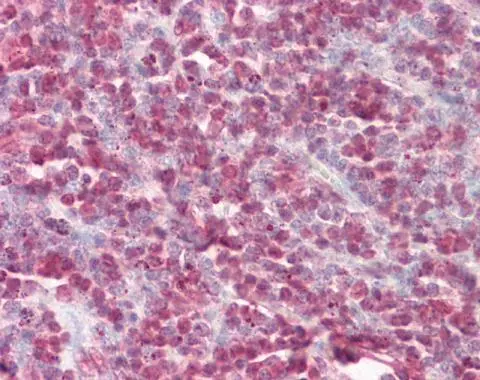
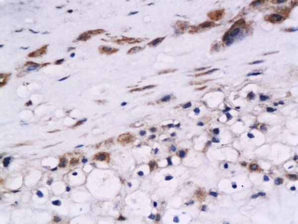
Cystatin B antibody detects Cystatin B protein at cytoplasm and nucleus human on tonsil by immunohistochemical analysis. Sample: Paraffin-embedded tonsil. Cystatin B antibody (GTX101458) dilution: 1:100.
Antigen Retrieval: Citrate buffer, pH 6.0, 15 min
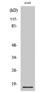
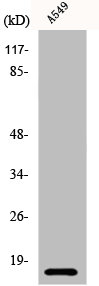
 diluted at 1:100.
Antigen Retrieval: Citrate buffer, pH 6.0, 15 min")
 of paraformaldehyde-fixed A431, using Cystatin B(GTX101458) antibody (Green) at 1:500 dilution. Alpha-tubulin filaments were labeled with GTX11304 (Red) at 1:2000.")
Cystatin B antibody detects Cystatin B protein at cytoplasm and nucleus human on tonsil by immunohistochemical analysis. Sample: Paraffin-embedded tonsil. Cystatin B antibody (GTX101458) dilution: 1:100.
Antigen Retrieval: Citrate buffer, pH 6.0, 15 min
Cystatin B antibody
GTX101458
ApplicationsImmunoFluorescence, ImmunoCytoChemistry, ImmunoHistoChemistry, ImmunoHistoChemistry Paraffin
Product group Antibodies
ReactivityHuman, Mouse
TargetCSTB
Overview
- SupplierGeneTex
- Product NameCystatin B antibody
- Delivery Days Customer9
- Application Supplier NoteICC/IF: 1:100-1:1000. IHC-P: 1:100-1:1000. *Optimal dilutions/concentrations should be determined by the researcher.Not tested in other applications.
- ApplicationsImmunoFluorescence, ImmunoCytoChemistry, ImmunoHistoChemistry, ImmunoHistoChemistry Paraffin
- CertificationResearch Use Only
- ClonalityPolyclonal
- Concentration6.04 mg/ml
- ConjugateUnconjugated
- Gene ID1476
- Target nameCSTB
- Target descriptioncystatin B
- Target synonymsCPI-B, CST6, EPM1, EPM1A, PME, STFB, ULD, cystatin-B, cystatin B (stefin B), epididymis secretory sperm binding protein, liver thiol proteinase inhibitor
- HostRabbit
- IsotypeIgG
- Protein IDP04080
- Protein NameCystatin-B
- Scientific DescriptionThe cystatin superfamily encompasses proteins that contain multiple cystatin-like sequences. Some of the members are active cysteine protease inhibitors, while others have lost or perhaps never acquired this inhibitory activity. There are three inhibitory families in the superfamily, including the type 1 cystatins (stefins), type 2 cystatins and kininogens. This gene encodes a stefin that functions as an intracellular thiol protease inhibitor. The protein is able to form a dimer stabilized by noncovalent forces, inhibiting papain and cathepsins l, h and b. The protein is thought to play a role in protecting against the proteases leaking from lysosomes. Evidence indicates that mutations in this gene are responsible for the primary defects in patients with progressive myoclonic epilepsy (EPM1). [provided by RefSeq]
- ReactivityHuman, Mouse
- Storage Instruction-20°C or -80°C,2°C to 8°C
- UNSPSC41116161
Datasheet
Related products
Product group Antibodies
Anti-Cystatin B AntibodyA34283
ApplicationsImmunoFluorescence, Western Blot, ELISA, ImmunoHistoChemistry
ReactivityHuman, Rat
- SizePrice

Product group Antibodies
Cystatin B Polyclonal AntibodyBS-5158R
ApplicationsImmunoFluorescence, ELISA, ImmunoCytoChemistry, ImmunoHistoChemistry, ImmunoHistoChemistry Frozen, ImmunoHistoChemistry Paraffin
ReactivityHuman, Mouse, Porcine, Rat
TargetCSTB
- SizePrice
Product group Antibodies
CSTB AntibodyCSB-PA002009
ApplicationsImmunoFluorescence, Western Blot, ELISA, ImmunoHistoChemistry
ReactivityHuman, Rat
TargetCSTB
- SizePrice
Product group Antibodies

ApplicationsWestern Blot, ELISA, ImmunoHistoChemistry
ReactivityHuman
TargetCSTB
- SizePrice
Product group Antibodies
Cstb Polyclonal AntibodyCAC08250
ApplicationsImmunoFluorescence, ImmunoPrecipitation, Western Blot, ELISA, ImmunoHistoChemistry
TargetCSTB
- SizePrice
Product group Antibodies

CSTB / Cystatin B / Stefin B AntibodyLS-C403094
ApplicationsELISA, ImmunoHistoChemistry
ReactivityHuman
TargetCSTB
- SizePrice
Product group Antibodies
Cystatin B antibody, C-termGTX15970
ApplicationsWestern Blot, ImmunoHistoChemistry, ImmunoHistoChemistry Paraffin
ReactivityHuman
TargetCSTB
- SizePrice
Product group Antibodies
Cystatin B antibodyGTX51631
ApplicationsImmunoHistoChemistry, ImmunoHistoChemistry Paraffin
ReactivityHuman
TargetCSTB
- SizePrice
Product group Antibodies
Cystatin B antibody [3F13]GTX52681
ApplicationsWestern Blot, Neutralisation/Blocking
ReactivityHuman
TargetCSTB
- SizePrice