

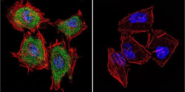
ICC/IF analysis of HeLa cells using GTX22727 Cytohesin 1 antibody [2E11]. Cells were probed without (right) or with(left) an antibody. Green : Primary antibody Blue : Nuclei Red : Actin Fixation : 4% paraformaldehyde Permeabilization : 0.1% Triton X-100 for 10 minute Dilution : 2 μg/ml in 0.1% BSA and incubated for 3 hours at room temperature
![ICC/IF analysis of HepG2 cells using GTX22727 Cytohesin 1 antibody [2E11]. Cells were probed without (right) or with(left) an antibody. Green : Primary antibody Blue : Nuclei Red : Actin Fixation : formaldehyde Dilution : 1:20 overnight at 4oC](https://www.genetex.com/upload/website/prouct_img/normal/GTX22727/GTX22727_387_ICC-IF_w_23060620_316.webp "ICC/IF analysis of HepG2 cells using GTX22727 Cytohesin 1 antibody [2E11]. Cells were probed without (right) or with(left) an antibody. Green : Primary antibody Blue : Nuclei Red : Actin Fixation : formaldehyde Dilution : 1:20 overnight at 4oC")
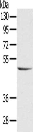
![WB analysis of whole cell extracts (30 μg lysate) of Raji (Lane 1), Jurkat (Lane 2) and Hep G2 (Lane 3) using GTX22727 Cytohesin 1 antibody [2E11]. Dilution : 0.5-2 μg/ml](https://www.genetex.com/upload/website/prouct_img/normal/GTX22727/GTX22727_1576_WB_w_23060620_570.webp "WB analysis of whole cell extracts (30 μg lysate) of Raji (Lane 1), Jurkat (Lane 2) and Hep G2 (Lane 3) using GTX22727 Cytohesin 1 antibody [2E11]. Dilution : 0.5-2 μg/ml")
![ICC/IF analysis of NIH-3T3 cells using GTX22727 Cytohesin 1 antibody [2E11]. Cells were probed without (right) or with(left) an antibody. Green : Primary antibody Blue : Nuclei Red : Actin Fixation : formaldehyde Dilution : 1:20 overnight at 4oC](https://www.genetex.com/upload/website/prouct_img/normal/GTX22727/GTX22727_385_ICC-IF_w_23060620_154.webp "ICC/IF analysis of NIH-3T3 cells using GTX22727 Cytohesin 1 antibody [2E11]. Cells were probed without (right) or with(left) an antibody. Green : Primary antibody Blue : Nuclei Red : Actin Fixation : formaldehyde Dilution : 1:20 overnight at 4oC")
![ICC/IF analysis of HeLa cells using GTX22727 Cytohesin 1 antibody [2E11]. Cells were probed without (right) or with(left) an antibody. Green : Primary antibody Blue : Nuclei Red : Actin Fixation : formaldehyde Dilution : 1:20 overnight at 4oC](https://www.genetex.com/upload/website/prouct_img/normal/GTX22727/GTX22727_386_ICC-IF_w_23060620_527.webp "ICC/IF analysis of HeLa cells using GTX22727 Cytohesin 1 antibody [2E11]. Cells were probed without (right) or with(left) an antibody. Green : Primary antibody Blue : Nuclei Red : Actin Fixation : formaldehyde Dilution : 1:20 overnight at 4oC")
ICC/IF analysis of HeLa cells using GTX22727 Cytohesin 1 antibody [2E11]. Cells were probed without (right) or with(left) an antibody. Green : Primary antibody Blue : Nuclei Red : Actin Fixation : 4% paraformaldehyde Permeabilization : 0.1% Triton X-100 for 10 minute Dilution : 2 μg/ml in 0.1% BSA and incubated for 3 hours at room temperature
Cytohesin 1 antibody [2E11]
GTX22727
ApplicationsImmunoFluorescence, Western Blot, ImmunoCytoChemistry
Product group Antibodies
ReactivityHuman, Mouse
TargetCYTH1
Overview
- SupplierGeneTex
- Product NameCytohesin 1 antibody [2E11]
- Delivery Days Customer9
- Application Supplier NoteWB: 1.0 microg/ml. ICC/IF: 1:20. *Optimal dilutions/concentrations should be determined by the researcher.Not tested in other applications.
- ApplicationsImmunoFluorescence, Western Blot, ImmunoCytoChemistry
- CertificationResearch Use Only
- ClonalityMonoclonal
- Concentration1 mg/ml
- ConjugateUnconjugated
- Gene ID9267
- Target nameCYTH1
- Target descriptioncytohesin 1
- Target synonymsB2-1, CYTOHESIN-1, D17S811E, PSCD1, SEC7, cytohesin-1, PH, SEC7 and coiled-coil domain-containing protein 1, SEC7 homolog B2-1, cytoadhesin 1, homolog of secretory protein SEC7, pleckstrin homology, Sec7 and coiled-coil domains 1
- HostMouse
- IsotypeIgG1
- Protein IDQ15438
- Protein NameCytohesin-1
- Scientific DescriptionThe protein encoded by this gene is a member of the PSCD family. Members of this family have identical structural organization that consists of an N-terminal coiled-coil motif, a central Sec7 domain, and a C-terminal pleckstrin homology (PH) domain. The coiled-coil motif is involved in homodimerization, the Sec7 domain contains guanine-nucleotide exchange protein activity, and the PH domain interacts with phospholipids and is responsible for association of PSCDs with membranes. Members of this family appear to mediate the regulation of protein sorting and membrane trafficking. This gene is highly expressed in natural killer and peripheral T cells, and regulates the adhesiveness of integrins at the plasma membrane of lymphocytes. A pseudogene of this gene has been defined on the X chromosome. Alternative splicing results in multiple transcript variants. [provided by RefSeq, May 2014]
- ReactivityHuman, Mouse
- Storage Instruction-20°C or -80°C,2°C to 8°C
- UNSPSC41116161
Datasheet
Related products
Product group Antibodies
CYTH1 AntibodyCSB-PA318480
ApplicationsWestern Blot, ELISA
ReactivityHuman, Mouse, Rat
TargetCYTH1
- SizePrice

Product group Antibodies
ApplicationsImmunoFluorescence, Western Blot, ImmunoCytoChemistry
ReactivityHuman, Mouse, Rat
TargetCYTH1
- SizePrice

Product group Antibodies
ApplicationsImmunoFluorescence, Western Blot, ImmunoCytoChemistry
ReactivityHuman, Mouse, Rat
- SizePrice
Product group Antibodies

CYTH1 / Cytohesin-1 AntibodyLS-C406702
ApplicationsWestern Blot, ELISA, ImmunoHistoChemistry
ReactivityHuman, Mouse, Rat
TargetCYTH1
- SizePrice
Product group Antibodies
ApplicationsFlow Cytometry
TargetCYTH1
- SizePrice
Product group Antibodies
Cytohesin 1 antibodyGTX66363
ApplicationsImmunoFluorescence, Western Blot, ImmunoCytoChemistry
ReactivityHuman, Mouse, Rat
TargetCYTH1
- SizePrice

Product group Antibodies
Cytohesin 1 antibody [N1C1]GTX102825
ApplicationsWestern Blot, ImmunoHistoChemistry
ReactivityHuman, Zebra Fish
TargetCYTH1
- SizePrice
Product group Antibodies
Cytohesin 1 antibody [CYT1-82]GTX12425
ApplicationsImmunoFluorescence, Western Blot, ELISA, ImmunoCytoChemistry
ReactivityHuman
TargetCYTH1
- SizePrice
Product group Antibodies
Anti-CYTH1 Antibody144-61008
ApplicationsWestern Blot
ReactivityHuman, Mouse, Rat
TargetCYTH1
- SizePrice