

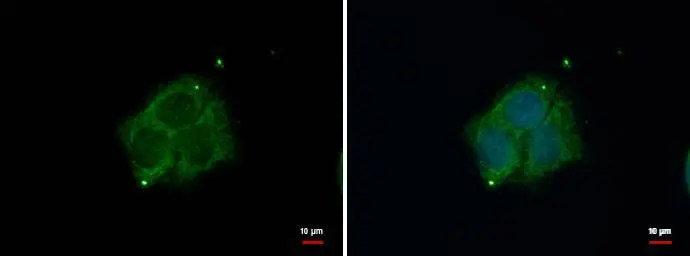
DAB1 antibody [N1N3] detects DAB1 protein at mitochondria by immunofluorescent analysis. Sample: HepG2 cells were fixed in 4% paraformaldehyde at RT for 15 min. Green: DAB1 protein stained by DAB1 antibody [N1N3] (GTX119336) diluted at 1:500. Blue: Hoechst 33342 staining.
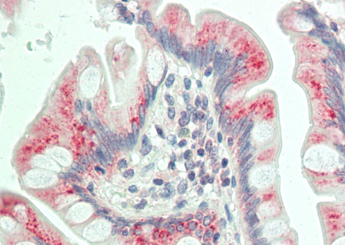
![DAB1 antibody [N1N3] detects DAB1 protein at cytosol on mouse duodenum by immunohistochemical analysis. Sample: Paraffin-embedded mouse duodenum. DAB1 antibody [N1N3] (GTX119336) dilution: 1:500.
Antigen Retrieval: Trilogy? (EDTA based, pH 8.0) buffer, 15min](https://www.genetex.com/upload/website/prouct_img/normal/GTX119336/GTX119336_40359_IHC_M_w_23060519_329.webp "DAB1 antibody [N1N3] detects DAB1 protein at cytosol on mouse duodenum by immunohistochemical analysis. Sample: Paraffin-embedded mouse duodenum. DAB1 antibody [N1N3] (GTX119336) dilution: 1:500.
Antigen Retrieval: Trilogy? (EDTA based, pH 8.0) buffer, 15min")
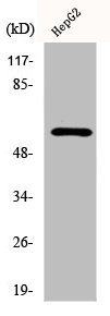
![Various tissue extracts (50 μg) were separated by 7.5% SDS-PAGE, and the membrane was blotted with Dab1 antibody [N1N3] (GTX119336) diluted at 1:1000. The HRP-conjugated anti-rabbit IgG antibody (GTX213110-01) was used to detect the primary antibody.](https://www.genetex.com/upload/website/prouct_img/normal/GTX119336/GTX119336_42228_20170504_WB_M_R_24022619_229.webp "Various tissue extracts (50 μg) were separated by 7.5% SDS-PAGE, and the membrane was blotted with Dab1 antibody [N1N3] (GTX119336) diluted at 1:1000. The HRP-conjugated anti-rabbit IgG antibody (GTX213110-01) was used to detect the primary antibody.")
![Various tissue extracts (50 μg) were separated by 7.5% SDS-PAGE, and the membrane was blotted with Dab1 antibody [N1N3] (GTX119336) diluted at 1:1000. The HRP-conjugated anti-rabbit IgG antibody (GTX213110-01) was used to detect the primary antibody. Corresponding RNA expression data are based on NCBI database.](https://www.genetex.com/upload/website/prouct_img/normal/GTX119336/GTX119336_42228_20250808_WB_R_tissue_RPKM_watermark_25081423_985.webp "Various tissue extracts (50 μg) were separated by 7.5% SDS-PAGE, and the membrane was blotted with Dab1 antibody [N1N3] (GTX119336) diluted at 1:1000. The HRP-conjugated anti-rabbit IgG antibody (GTX213110-01) was used to detect the primary antibody. Corresponding RNA expression data are based on NCBI database.")
DAB1 antibody [N1N3] detects DAB1 protein at mitochondria by immunofluorescent analysis. Sample: HepG2 cells were fixed in 4% paraformaldehyde at RT for 15 min. Green: DAB1 protein stained by DAB1 antibody [N1N3] (GTX119336) diluted at 1:500. Blue: Hoechst 33342 staining.
Dab1 antibody [N1N3]
GTX119336
ApplicationsImmunoFluorescence, Western Blot, ImmunoCytoChemistry, ImmunoHistoChemistry, ImmunoHistoChemistry Paraffin
Product group Antibodies
ReactivityHuman, Mouse, Rat
TargetDAB1
Overview
- SupplierGeneTex
- Product NameDab1 antibody [N1N3]
- Delivery Days Customer9
- Application Supplier NoteWB: 1:500-1:3000. ICC/IF: 1:100-1:1000. IHC-P: 1:100-1:1000. *Optimal dilutions/concentrations should be determined by the researcher.Not tested in other applications.
- ApplicationsImmunoFluorescence, Western Blot, ImmunoCytoChemistry, ImmunoHistoChemistry, ImmunoHistoChemistry Paraffin
- CertificationResearch Use Only
- ClonalityPolyclonal
- Concentration0.73 mg/ml
- ConjugateUnconjugated
- Gene ID1600
- Target nameDAB1
- Target descriptionDAB adaptor protein 1
- Target synonymsSCA37, disabled homolog 1, DAB1, reelin adaptor protein, Dab reelin signal transducer 1, Dab, reelin signal transducer, homolog 1, spinocerebellar ataxia 37, yotari
- HostRabbit
- IsotypeIgG
- Protein IDO75553
- Protein NameDisabled homolog 1
- Scientific DescriptionThe laminar organization of multiple neuronal types in the cerebral cortex is required for normal cognitive function. In mice, the disabled-1 gene plays a central role in brain development, directing the migration of cortical neurons past previously formed neurons to reach their proper layer. This gene is similar to disabled-1, and the protein encoded by this gene is thought to be a signal transducer that interacts with protein kinase pathways to regulate neuronal positioning in the developing brain. Alternatively spliced transcript variants of this gene have been reported, but their full length nature has not been determined. [provided by RefSeq]
- ReactivityHuman, Mouse, Rat
- Storage Instruction-20°C or -80°C,2°C to 8°C
- UNSPSC41116161
Datasheet
Related products
Product group Antibodies
ApplicationsImmunoFluorescence, Western Blot, ImmunoHistoChemistry
ReactivityHuman, Mouse, Rat
- SizePrice
Product group Antibodies
Anti-Phospho-Dab1 (Y232) AntibodyA03459Y232
ApplicationsImmunoFluorescence, Western Blot, ELISA
ReactivityHuman, Mouse, Rat
TargetDAB1
- SizePrice
Product group Antibodies
Anti-DAB1 Antibody144-61843
ApplicationsWestern Blot
ReactivityHuman, Mouse
TargetDAB1
- SizePrice
Product group Antibodies
Dab1 Polyclonal AntibodyBS-11547R
ApplicationsImmunoFluorescence, ELISA, ImmunoCytoChemistry, ImmunoHistoChemistry, ImmunoHistoChemistry Frozen, ImmunoHistoChemistry Paraffin
ReactivityBovine, Canine, Equine, Human, Mouse, Porcine, Rabbit, Rat
- SizePrice
Product group Antibodies
DAB1 AntibodyCSB-PA002060
ApplicationsImmunoFluorescence, Western Blot, ELISA
ReactivityHuman, Mouse, Rat
TargetDAB1
- SizePrice
Product group Antibodies

DAB1 AntibodyLS-C497058
ApplicationsWestern Blot
ReactivityHuman, Mouse
TargetDAB1
- SizePrice

Product group Antibodies
Anti-DAB1 AntibodyHPA052033
ApplicationsImmunoCytoChemistry, ImmunoHistoChemistry
ReactivityHuman, Mouse
TargetDAB1
- SizePrice