

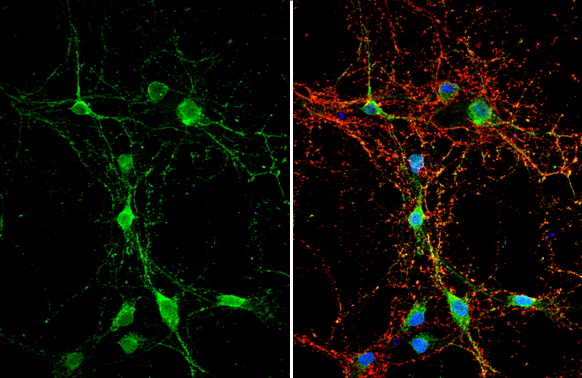
DAG1 antibody detects DAG1 protein by immunofluorescent analysis. Sample: DIV10 rat E18 primary hippocampal neuron cells were fixed in 4% paraformaldehyde at RT for 15 min. Green: DAG1 stained by DAG1 antibody (GTX105038) diluted at 1:500. Red: Tau, stained by Tau antibody [GT287] (GTX634809) diluted at 1:500. Blue: Fluoroshield with DAPI (GTX30920).
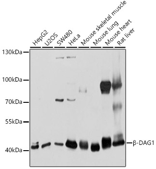
 were separated by 7.5% SDS-PAGE, and the membrane was blotted with DAG1 antibody (GTX105038) diluted at 1:500. The HRP-conjugated anti-rabbit IgG antibody (GTX213110-01) was used to detect the primary antibody.")
 were separated by 7.5% SDS-PAGE, and the membrane was blotted with DAG1 antibody (GTX105038) diluted at 1:500. The HRP-conjugated anti-rabbit IgG antibody (GTX213110-01) was used to detect the primary antibody.")
 were separated by 7.5% SDS-PAGE, and the membrane was blotted with DAG1 antibody (GTX105038) diluted at 1:500. The HRP-conjugated anti-rabbit IgG antibody (GTX213110-01) was used to detect the primary antibody.")
![alpha Dystroglycan antibody detects alpha Dystroglycan protein at cytoplasm by immunofluorescent analysis. Sample: HeLa cells were fixed in 4% paraformaldehyde at RT for 15 min. Green: alpha Dystroglycan protein stained by alpha Dystroglycan antibody (GTX105038) diluted at 1:1000. Red: alpha Tubulin, a cytoskeleton marker, stained by alpha Tubulin antibody [B-5-1-2] (GTX11304) diluted at 1:10000. Blue: Hoechst 33342 staining.](https://www.genetex.com/upload/website/prouct_img/normal/GTX105038/GTX105038_39876_20150410_IFA_w_23060120_405.webp "alpha Dystroglycan antibody detects alpha Dystroglycan protein at cytoplasm by immunofluorescent analysis. Sample: HeLa cells were fixed in 4% paraformaldehyde at RT for 15 min. Green: alpha Dystroglycan protein stained by alpha Dystroglycan antibody (GTX105038) diluted at 1:1000. Red: alpha Tubulin, a cytoskeleton marker, stained by alpha Tubulin antibody [B-5-1-2] (GTX11304) diluted at 1:10000. Blue: Hoechst 33342 staining.")
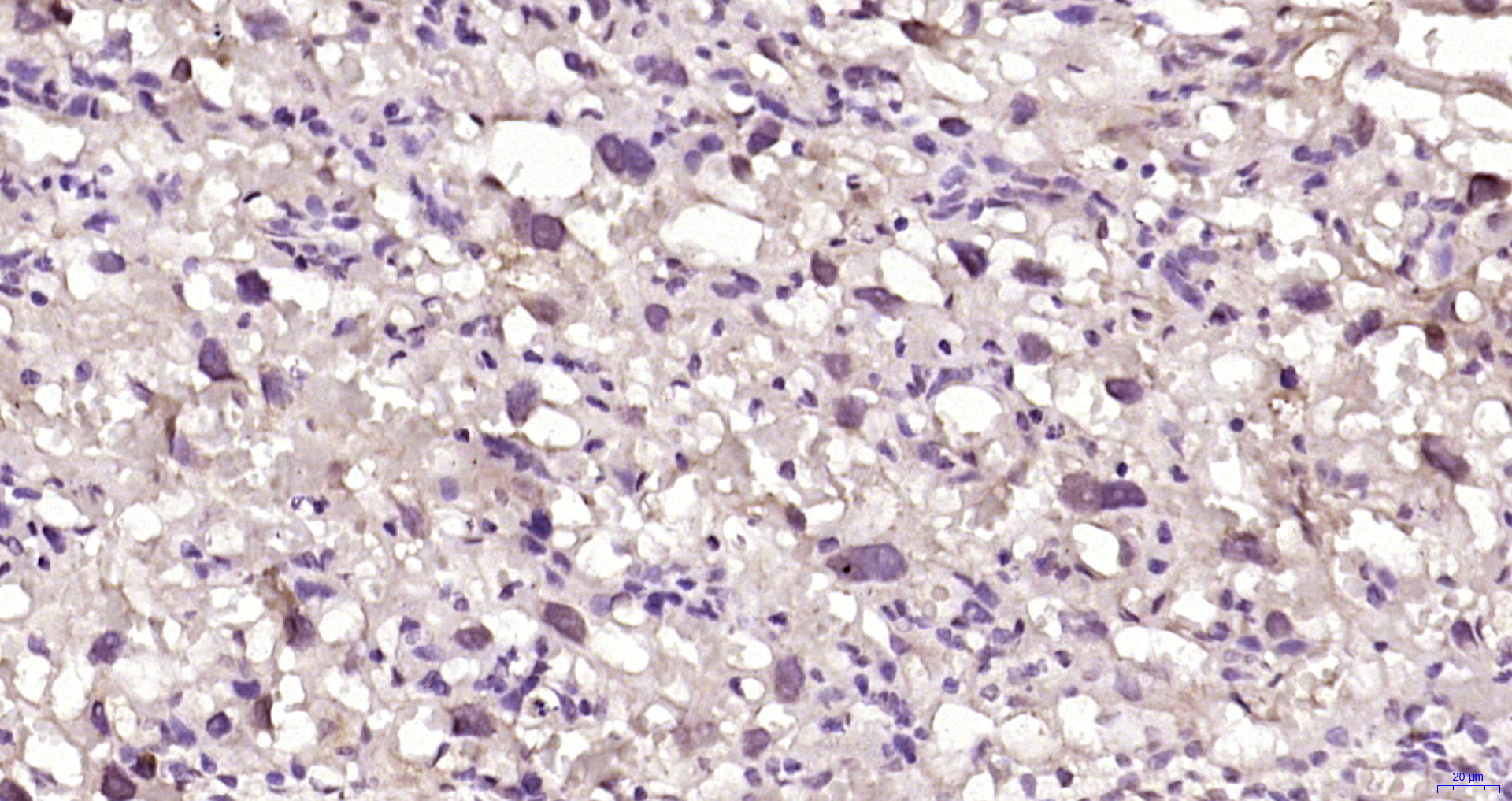
 diluted at 1:500. Antigen Retrieval: Citrate buffer, pH 6.0, 15 min")
 were separated by 7.5% SDS-PAGE, and the membrane was blotted with DAG1 antibody (GTX105038) diluted at 1:500. The HRP-conjugated anti-rabbit IgG antibody (GTX213110-01) was used to detect the primary antibody, and the signal was developed with Trident ECL plus-Enhanced.")
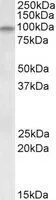
 was separated by 7.5% SDS-PAGE, and the membrane was blotted with DAG1 antibody (GTX105038) diluted at 1:1000. The HRP-conjugated anti-rabbit IgG antibody (GTX213110-01) was used to detect the primary antibody.")
DAG1 antibody detects DAG1 protein by immunofluorescent analysis. Sample: DIV10 rat E18 primary hippocampal neuron cells were fixed in 4% paraformaldehyde at RT for 15 min. Green: DAG1 stained by DAG1 antibody (GTX105038) diluted at 1:500. Red: Tau, stained by Tau antibody [GT287] (GTX634809) diluted at 1:500. Blue: Fluoroshield with DAPI (GTX30920).
DAG1 antibody
GTX105038
ApplicationsImmunoFluorescence, Western Blot, ImmunoCytoChemistry, ImmunoHistoChemistry, ImmunoHistoChemistry Paraffin
Product group Antibodies
ReactivityEquine, Human, Mouse, Rat
TargetDAG1
Overview
- SupplierGeneTex
- Product NameDAG1 antibody
- Delivery Days Customer9
- Application Supplier NoteWB: 1:500-1:3000. ICC/IF: 1:100-1:1000. IHC-P: 1:100-1:1000. *Optimal dilutions/concentrations should be determined by the researcher.Not tested in other applications.
- ApplicationsImmunoFluorescence, Western Blot, ImmunoCytoChemistry, ImmunoHistoChemistry, ImmunoHistoChemistry Paraffin
- CertificationResearch Use Only
- ClonalityPolyclonal
- Concentration1.28 mg/ml
- ConjugateUnconjugated
- Gene ID1605
- Target nameDAG1
- Target descriptiondystroglycan 1
- Target synonyms156DAG, A3a, AGRNR, DAG, LGMDR16, MDDGA9, MDDGC7, MDDGC9, dystroglycan 1, dystroglycan 1 (dystrophin-associated glycoprotein 1)
- HostRabbit
- IsotypeIgG
- Protein IDQ14118
- Protein NameDystroglycan 1
- Scientific DescriptionDystroglycan is a laminin binding component of the dystrophin-glycoprotein complex which provides a linkage between the subsarcolemmal cytoskeleton and the extracellular matrix. Dystroglycan 1 is a candidate gene for the site of the mutation in autosomal recessive muscular dystrophies. The dramatic reduction of dystroglycan 1 in Duchenne muscular dystrophy leads to a loss of linkage between the sarcolemma and extracellular matrix, rendering muscle fibers more susceptible to necrosis. Dystroglycan also functions as dual receptor for agrin and laminin-2 in the Schwann cell membrane. The muscle and nonmuscle isoforms of dystroglycan differ by carbohydrate moieties but not protein sequence. [provided by RefSeq]
- ReactivityEquine, Human, Mouse, Rat
- Storage Instruction-20°C or -80°C,2°C to 8°C
- UNSPSC41116161
Datasheet
Related products
Product group Antibodies

Anti-Beta Dystroglycan [AS30SS_Hu6]Ab03277-10.0
ApplicationsImmunoFluorescence, ImmunoPrecipitation, Western Blot, ELISA
ReactivityHuman, Mouse
TargetDAG1
- SizePrice
Product group Antibodies
Anti-DAG1 AntibodyA12781
ApplicationsImmunoFluorescence, ImmunoPrecipitation, Western Blot, ImmunoCytoChemistry
ReactivityHuman, Mouse, Rat
- SizePrice

Product group Antibodies
ApplicationsImmunoFluorescence, ImmunoPrecipitation, Western Blot, ImmunoCytoChemistry
ReactivityHuman, Mouse, Rat
TargetDAG1
- SizePrice
Product group Antibodies
Anti-DAG1 Antibody144-10076
ApplicationsWestern Blot
ReactivityHuman, Mouse, Rat
TargetDAG1
- SizePrice
Product group Antibodies
ApplicationsImmunoFluorescence, Western Blot, ELISA, ImmunoCytoChemistry, ImmunoHistoChemistry, ImmunoHistoChemistry Frozen, ImmunoHistoChemistry Paraffin
ReactivityBovine, Equine, Guinea Pig, Human, Mouse, Porcine, Rabbit, Rat
TargetDAG1
- SizePrice
Product group Antibodies
DAG1 AntibodyCSB-PA613486NA01HU
ApplicationsELISA, ImmunoHistoChemistry
ReactivityHuman
TargetDAG1
- SizePrice
![IHC-Fr analysis of human skeletal muscle using GTX01940 DAG1 / beta Dystroglycan antibody [43DAG1/8D5]. Staining is localized in the sarcolemma of the fibers.](https://www.genetex.com/upload/website/prouct_img/normal/GTX01940/GTX01940_20200811_IHC-Fr_106_w_23053121_152.webp)
Product group Antibodies
ApplicationsImmunoHistoChemistry, ImmunoHistoChemistry Frozen
ReactivityAmphibian, Canine, Chicken, Hamster, Human, Mouse, Rabbit, Rat
TargetDAG1
- SizePrice
Product group Antibodies

DAG1 / Dystroglycan AntibodyLS-C496786
ApplicationsWestern Blot
ReactivityHuman, Mouse, Rat
TargetDAG1
- SizePrice
Product group Antibodies
DAG1 antibody, InternalGTX88089
ApplicationsWestern Blot
ReactivityHuman, Mouse, Rat
TargetDAG1
- SizePrice

Product group Antibodies
Anti-DAG1 AntibodyHPA070563
ApplicationsImmunoCytoChemistry
ReactivityHuman
TargetDAG1
- SizePrice