

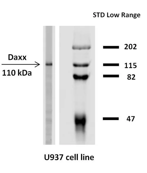
WB analysis of U937 cell lysate using GTX80185 Daxx antibody [DAXX-03].
WB analysis of U937 cell lysate using GTX80185 Daxx antibody [DAXX-03].
Daxx antibody [DAXX-03]
GTX80185
ApplicationsImmunoFluorescence, ImmunoPrecipitation, Western Blot, ImmunoCytoChemistry
Product group Antibodies
ReactivityHuman
TargetDAXX
Overview
- SupplierGeneTex
- Product NameDaxx antibody [DAXX-03]
- Delivery Days Customer9
- ApplicationsImmunoFluorescence, ImmunoPrecipitation, Western Blot, ImmunoCytoChemistry
- CertificationResearch Use Only
- ClonalityMonoclonal
- Clone IDDAXX-03
- Concentration1 mg/ml
- ConjugateUnconjugated
- Gene ID1616
- Target nameDAXX
- Target descriptiondeath domain associated protein
- Target synonymsBING2, DAP6, EAP1, SMIM40, death domain-associated protein 6, CENP-C binding protein, ETS1-associated protein 1, Fas-binding protein, death-associated protein 6, fas death domain-associated protein
- HostMouse
- IsotypeIgG1
- Protein IDQ9UER7
- Protein NameDeath domain-associated protein 6
- Scientific DescriptionThis gene encodes a multifunctional protein that resides in multiple locations in the nucleus and in the cytoplasm. It interacts with a wide variety of proteins, such as apoptosis antigen Fas, centromere protein C, and transcription factor erythroblastosis virus E26 oncogene homolog 1. In the nucleus, the encoded protein functions as a potent transcription repressor that binds to sumoylated transcription factors. Its repression can be relieved by the sequestration of this protein into promyelocytic leukemia nuclear bodies or nucleoli. This protein also associates with centromeres in G2 phase. In the cytoplasm, the encoded protein may function to regulate apoptosis. The subcellular localization and function of this protein are modulated by post-translational modifications, including sumoylation, phosphorylation and polyubiquitination. Alternative splicing results in multiple transcript variants. [provided by RefSeq, Nov 2008]
- ReactivityHuman
- Storage Instruction2°C to 8°C
- UNSPSC41116161
Datasheet
Related products
Product group Antibodies
DAXX AntibodyCSB-PA002077
ApplicationsImmunoFluorescence, Western Blot, ELISA, ImmunoHistoChemistry
ReactivityHuman, Mouse, Rat
TargetDAXX
- SizePrice
Product group Antibodies
Anti-DAXX AntibodyA96404
ApplicationsImmunoFluorescence, Western Blot, ELISA
ReactivityHuman, Mouse, Rat
- SizePrice

Product group Antibodies
Anti-DAXX AntibodyAMAB91191
ApplicationsImmunoCytoChemistry, ImmunoHistoChemistry
ReactivityHuman
TargetDAXX
- SizePrice
Product group Antibodies

DAXX Antibody (aa466-516)LS-C288166
ApplicationsWestern Blot
ReactivityHuman
TargetDAXX
- SizePrice
![ICC/IF analysis of HeLa cells transfected with Myc-Daxx only (Panel A) or co-transfected with MycDaxx / ASK1HA (Panel B) using GTX29091 Daxx antibody [DAXX-01]. Co-transfected with pCDNA3-MycDaxx and pCDNA3-ASK1HA, which led to translocation of Daxx from nucleus to cytoplasm. Green : Primary antibody Blue : DAPI](https://www.genetex.com/upload/website/prouct_img/normal/GTX29091/GTX29091_20191028_ICC-IF_1_w_23060722_949.webp)
Product group Antibodies
Daxx antibody [DAXX-01]GTX29091
ApplicationsImmunoFluorescence, Western Blot, ImmunoCytoChemistry
ReactivityHuman
TargetDAXX
- SizePrice

Product group Antibodies
Anti-Daxx Antibody Picoband(r)PB9550-CARRIER-FREE
ApplicationsFlow Cytometry, ImmunoFluorescence, Western Blot, ImmunoCytoChemistry, ImmunoHistoChemistry
ReactivityHuman, Mouse, Rat
TargetDAXX
- SizePrice
Product group Antibodies
ApplicationsImmunoFluorescence, Western Blot, ELISA, ImmunoCytoChemistry, ImmunoHistoChemistry, ImmunoHistoChemistry Frozen, ImmunoHistoChemistry Paraffin
ReactivityBovine, Human, Porcine
TargetDAXX
- SizePrice
![ICC/IF analysis of PANC-1 cells using GTX83312 Daxx antibody [7A11]. Green : Daxx Blue: DRAQ5 fluorescent DNA dye](https://www.genetex.com/upload/website/prouct_img/normal/GTX83312/GTX83312_20170912_ICCIF_1_w_23061322_647.webp)
Product group Antibodies
Daxx antibody [7A11]GTX83312
ApplicationsFlow Cytometry, ImmunoFluorescence, Western Blot, ELISA, ImmunoCytoChemistry
ReactivityHuman
TargetDAXX
- SizePrice