

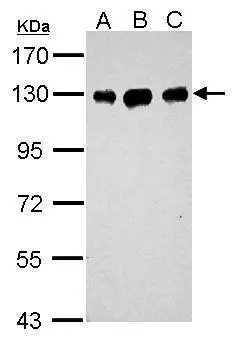
Sample (30 μg of whole cell lysate) A: Neuro2A B: GL261 C: C8D30 7.5% SDS PAGE GTX100232 diluted at 1:1000 The HRP-conjugated anti-rabbit IgG antibody (GTX213110-01) was used to detect the primary antibody.
 antibody at 1:500 dilution.
Antigen Retrieval: Trilogy? (EDTA based, pH 8.0) buffer, 15min")
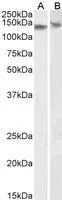
![Non-transfected (–) and transfected (+) 293T whole cell extracts (30 μg) were separated by 7.5% SDS-PAGE, and the membrane was blotted with DDB1 antibody [C3], C-term (GTX100232) diluted at 1:10000. The HRP-conjugated anti-rabbit IgG antibody (GTX213110-01) was used to detect the primary antibody.](https://www.genetex.com/upload/website/prouct_img/normal/GTX100232/GTX100232_40877_20160728_WB_shRNA_watermark_w_23053123_355.webp "Non-transfected (–) and transfected (+) 293T whole cell extracts (30 μg) were separated by 7.5% SDS-PAGE, and the membrane was blotted with DDB1 antibody [C3], C-term (GTX100232) diluted at 1:10000. The HRP-conjugated anti-rabbit IgG antibody (GTX213110-01) was used to detect the primary antibody.")
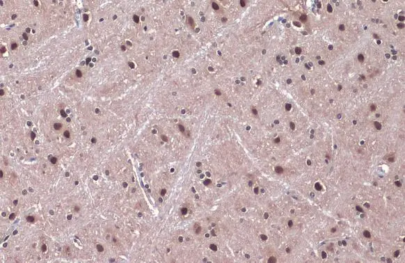
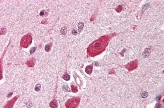
![DDB1 antibody [C3], C-term detects DDB1 protein at cytosol and nucleus on mouse hind brain by immunohistochemical analysis. Sample: Paraffin-embedded mouse hind brain. DDB1 antibody [C3], C-term (GTX100232) dilution: 1:500.
Antigen Retrieval: Trilogy? (EDTA based, pH 8.0) buffer, 15min](https://www.genetex.com/upload/website/prouct_img/normal/GTX100232/GTX100232_40877_IHC_M_2_w_23053123_865.webp "DDB1 antibody [C3], C-term detects DDB1 protein at cytosol and nucleus on mouse hind brain by immunohistochemical analysis. Sample: Paraffin-embedded mouse hind brain. DDB1 antibody [C3], C-term (GTX100232) dilution: 1:500.
Antigen Retrieval: Trilogy? (EDTA based, pH 8.0) buffer, 15min")
 A: PC-12 7.5% SDS PAGE GTX100232 diluted at 1:1000 The HRP-conjugated anti-rabbit IgG antibody (GTX213110-01) was used to detect the primary antibody.")
![DDB1 antibody [C3], C-term detects DDB1 protein at cytoplasm and nucleus by immunofluorescent analysis. Sample: HeLa cells were fixed in ice-cold MeOH for 5 min. Green: DDB1 protein stained by DDB1 antibody [C3], C-term (GTX100232) diluted at 1:500. Blue: Hoechst 33343 staining.](https://www.genetex.com/upload/website/prouct_img/normal/GTX100232/GTX100232_40877_IFA_w_23053123_101.webp "DDB1 antibody [C3], C-term detects DDB1 protein at cytoplasm and nucleus by immunofluorescent analysis. Sample: HeLa cells were fixed in ice-cold MeOH for 5 min. Green: DDB1 protein stained by DDB1 antibody [C3], C-term (GTX100232) diluted at 1:500. Blue: Hoechst 33343 staining.")
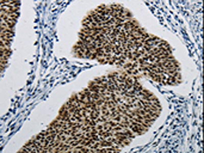
![DDB1 antibody [C3], C-term detects DDB1 protein at cytosol and nucleus on mouse duodenum by immunohistochemical analysis. Sample: Paraffin-embedded mouse duodenum. DDB1 antibody [C3], C-term (GTX100232) dilution: 1:500.
Antigen Retrieval: Trilogy? (EDTA based, pH 8.0) buffer, 15min](https://www.genetex.com/upload/website/prouct_img/normal/GTX100232/GTX100232_40877_IHC_M_w_23053123_255.webp "DDB1 antibody [C3], C-term detects DDB1 protein at cytosol and nucleus on mouse duodenum by immunohistochemical analysis. Sample: Paraffin-embedded mouse duodenum. DDB1 antibody [C3], C-term (GTX100232) dilution: 1:500.
Antigen Retrieval: Trilogy? (EDTA based, pH 8.0) buffer, 15min")
 A: A549 B: H1299 C: HCT116 D: MCF-7 7.5% SDS PAGE GTX100232 diluted at 1:1000 The HRP-conjugated anti-rabbit IgG antibody (GTX213110-01) was used to detect the primary antibody.")
![DDB1 antibody [C3], C-term detects DDB1 protein at cytosol and nucleus on mouse heart by immunohistochemical analysis. Sample: Paraffin-embedded mouse heart. DDB1 antibody [C3], C-term (GTX100232) dilution: 1:500.
Antigen Retrieval: Trilogy? (EDTA based, pH 8.0) buffer, 15min](https://www.genetex.com/upload/website/prouct_img/normal/GTX100232/GTX100232_40877_IHC_M_3_w_23053123_748.webp "DDB1 antibody [C3], C-term detects DDB1 protein at cytosol and nucleus on mouse heart by immunohistochemical analysis. Sample: Paraffin-embedded mouse heart. DDB1 antibody [C3], C-term (GTX100232) dilution: 1:500.
Antigen Retrieval: Trilogy? (EDTA based, pH 8.0) buffer, 15min")

Sample (30 μg of whole cell lysate) A: Neuro2A B: GL261 C: C8D30 7.5% SDS PAGE GTX100232 diluted at 1:1000 The HRP-conjugated anti-rabbit IgG antibody (GTX213110-01) was used to detect the primary antibody.
DDB1 antibody [C3], C-term
GTX100232
ApplicationsImmunoFluorescence, Western Blot, ImmunoCytoChemistry, ImmunoHistoChemistry, ImmunoHistoChemistry Paraffin
Product group Antibodies
ReactivityHuman, Mouse, Rat
TargetDDB1
Overview
- SupplierGeneTex
- Product NameDDB1 antibody [C3], C-term
- Delivery Days Customer9
- Application Supplier NoteWB: 1:500-1:10000. ICC/IF: 1:100-1:1000. IHC-P: 1:100-1:1000. *Optimal dilutions/concentrations should be determined by the researcher.Not tested in other applications.
- ApplicationsImmunoFluorescence, Western Blot, ImmunoCytoChemistry, ImmunoHistoChemistry, ImmunoHistoChemistry Paraffin
- CertificationResearch Use Only
- ClonalityPolyclonal
- Concentration1 mg/ml
- ConjugateUnconjugated
- Gene ID1642
- Target nameDDB1
- Target descriptiondamage specific DNA binding protein 1
- Target synonymsDDBA, UV-DDB1, WHIKERS, XAP1, XPCE, XPE, XPE-BF, DNA damage-binding protein 1, DDB p127 subunit, DNA damage-binding protein a, HBV X-associated protein 1, UV-DDB 1, UV-damaged DNA-binding factor, UV-damaged DNA-binding protein 1, XAP-1, XPE-binding factor, damage-specific DNA binding protein 1, 127kDa, xeroderma pigmentosum group E-complementing protein
- HostRabbit
- IsotypeIgG
- Protein IDQ16531
- Protein NameDNA damage-binding protein 1
- Scientific DescriptionThis gene encodes the large subunit of DNA damage-binding protein which is a heterodimer composed of a large and a small subunit. This protein functions in nucleotide-excision repair. Its defective activity causes the repair defect in the patients with xeroderma pigmentosum complementation group E (XPE). However, it remains for mutation analysis to demonstrate whether the defect in XPE patients is in this gene or the gene encoding the small subunit. In addition, Best vitelliform mascular dystrophy is mapped to the same region as this gene on 11q, but no sequence alternations of this gene are demonstrated in Best disease patients. [provided by RefSeq]
- ReactivityHuman, Mouse, Rat
- Storage Instruction-20°C or -80°C,2°C to 8°C
- UNSPSC41116161
Datasheet
Related products
Product group Antibodies
Anti-DDB1 AntibodyA30243
ApplicationsWestern Blot, ImmunoHistoChemistry
ReactivityHuman, Mouse, Rat
- SizePrice
Product group Antibodies
Anti-DDB1 Antibody144-02896
ApplicationsWestern Blot, ImmunoHistoChemistry
ReactivityHuman, Mouse, Rat
TargetDDB1
- SizePrice
Product group Antibodies
DDB1 Polyclonal AntibodyBS-2588R
ApplicationsImmunoFluorescence, ELISA, ImmunoCytoChemistry, ImmunoHistoChemistry, ImmunoHistoChemistry Frozen, ImmunoHistoChemistry Paraffin
ReactivityBovine, Chicken, Equine, Human, Mouse, Rabbit, Rat
TargetDDB1
- SizePrice
Product group Antibodies
References

Goat anti-DDB1EB05033
ApplicationsWestern Blot, ELISA, ImmunoHistoChemistry
ReactivityBovine, Canine, Human, Mouse, Rat
TargetDDB1
- SizePrice
Product group Antibodies
DDB1 AntibodyCSB-PA056628
ApplicationsWestern Blot, ELISA, ImmunoHistoChemistry
ReactivityHuman, Mouse, Rat
TargetDDB1
- SizePrice
Product group Antibodies

DDB1 AntibodyLS-C402236
ApplicationsWestern Blot, ELISA, ImmunoHistoChemistry
ReactivityHuman, Mouse, Rat
TargetDDB1
- SizePrice
![Non-transfected (–) and transfected (+) 293T whole cell extracts (30 μg) were separated by 7.5% SDS-PAGE, and the membrane was blotted with DDB1 antibody [N1N3] (GTX100129) diluted at 1:5000.](https://www.genetex.com/upload/website/prouct_img/normal/GTX100129/GTX100129_39384_20160728_WB_shRNA_watermark_w_23053123_216.webp)
Product group Antibodies
DDB1 antibody [N1N3]GTX100129
ApplicationsImmunoFluorescence, Western Blot, ImmunoCytoChemistry
ReactivityHuman, Mouse
TargetDDB1
- SizePrice
Product group Antibodies
DDB1 antibodyGTX100130
ApplicationsImmunoFluorescence, ImmunoPrecipitation, Western Blot, ImmunoCytoChemistry, ImmunoHistoChemistry, ImmunoHistoChemistry Paraffin
ReactivityHuman, Mouse, Rat
TargetDDB1
- SizePrice
Product group Antibodies
DDB1 antibodyGTX85639
ApplicationsImmunoPrecipitation, Western Blot, ELISA, ImmunoHistoChemistry, ImmunoHistoChemistry Paraffin
ReactivityHuman
TargetDDB1
- SizePrice
Product group Antibodies
DDB1 antibody, C-termGTX89975
ApplicationsWestern Blot
ReactivityHuman, Mouse
TargetDDB1
- SizePrice