

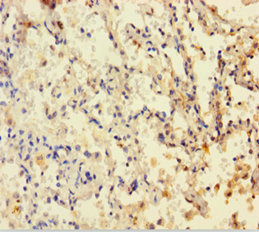
Immunohistochemistry of paraffin-embedded human lung cancer using CSB-PA006847HA01HU at dilution of 1:100
")
Immunohistochemistry of paraffin-embedded human lung cancer using CSB-PA006847HA01HU at dilution of 1:100
DHFR Antibody
CSB-PA006847HA01HU
ApplicationsImmunoFluorescence, ELISA, ImmunoHistoChemistry
Product group Antibodies
ReactivityHuman
TargetDHFR
Overview
- SupplierCusabio
- Product NameDHFR Antibody
- Delivery Days Customer20
- ApplicationsImmunoFluorescence, ELISA, ImmunoHistoChemistry
- CertificationResearch Use Only
- ClonalityPolyclonal
- ConjugateUnconjugated
- Gene ID1719
- Target nameDHFR
- Target descriptiondihydrofolate reductase
- Target synonymsDHFR1, DHFRP1, DYR, dihydrofolate reductase
- HostRabbit
- IsotypeIgG
- Protein IDP00374
- Protein NameDihydrofolate reductase
- Scientific DescriptionKey enzyme in folate metabolism. Contributes to the de novo mitochondrial thymidylate biosynthesis pathway. Catalyzes an essential reaction for de novo glycine and purine synthesis, and for DNA precursor synthesis. Binds its own mRNA and that of DHFRL1.
- ReactivityHuman
- Storage Instruction-20°C or -80°C
- UNSPSC41116161
Related products
Product group Antibodies
Anti-DHFR AntibodyA29840
ApplicationsWestern Blot, ImmunoHistoChemistry
ReactivityHuman, Mouse, Rat
- SizePrice
Product group Antibodies
Anti-DHFR Antibody144-01607
ApplicationsImmunoFluorescence, Western Blot
ReactivityHuman, Mouse
TargetDHFR
- SizePrice
Product group Antibodies

DHFR AntibodyLS-C830690
ApplicationsELISA, ImmunoHistoChemistry
ReactivityHuman, Mouse, Rat
TargetDHFR
- SizePrice
Product group Antibodies
DHFR Recombinant Antibody, AbBy Fluor-350 ConjugatedBSM-61547R-BF350
ApplicationsImmunoFluorescence, Western Blot
ReactivityHuman, Mouse, Rat
TargetDHFR
- SizePrice
Product group Antibodies
Dhfr Polyclonal AntibodyCAC11829
ApplicationsImmunoFluorescence, ELISA, ImmunoHistoChemistry
TargetDHFR
- SizePrice
Product group Antibodies
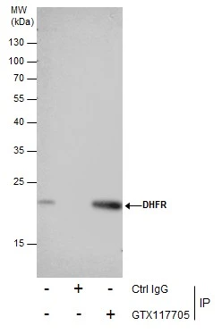
DHFR antibodyGTX117705
ApplicationsImmunoFluorescence, ImmunoPrecipitation, Western Blot, ImmunoCytoChemistry, ImmunoHistoChemistry, ImmunoHistoChemistry Paraffin
ReactivityHuman, Rat
TargetDHFR
- SizePrice
Product group Antibodies
Anti-SNRPD3 AntibodyCAB16070
ApplicationsWestern Blot, ELISA
ReactivityHuman
TargetDHFR
- SizePrice

Product group Antibodies
Anti-Dihydrofolate reductase (DHFR) Antibody Picoband(r)PB9175-CARRIER-FREE
ApplicationsFlow Cytometry, ImmunoFluorescence, Western Blot, ImmunoCytoChemistry, ImmunoHistoChemistry
ReactivityHuman, Mouse, Rat
TargetDHFR
- SizePrice