

DHFR Antibody (YA3335)
HY-P83590
TargetDHFR
Product group Antibodies
Overview
- SupplierMedChem Express
- Product NameDHFR Antibody (YA3335)
- Delivery Days Customer5
- CertificationResearch Use Only
- ClonalityMonoclonal
- Gene ID1719
- Target nameDHFR
- Target descriptiondihydrofolate reductase
- Target synonymsDHFRP1; dihydrofolate reductase; DYR
- HostRabbit
- IsotypeIgG
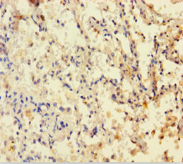
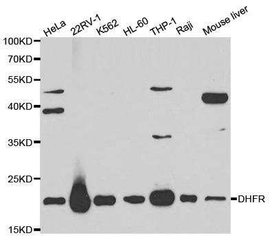
- Scientific DescriptionDHFR Antibody (YA3335) is a rabbit-derived non-conjugated IgG antibody (Clone NO.: YA3335), targeting DHFR, with a predicted molecular weight of 21 kDa (observed band size: 21 kDa). DHFR Antibody (YA3335) can be used for WB, IHC-P, ICC/IF experiment in human, mouse, rat background.
- UNSPSC12352203
Related products
Product group Antibodies
Dhfr Polyclonal AntibodyCAC11829
ApplicationsImmunoFluorescence, ELISA, ImmunoHistoChemistry
TargetDHFR
- SizePrice
Product group Antibodies
References
DHFR antibodyGTX117705
ApplicationsImmunoFluorescence, ImmunoPrecipitation, Western Blot, ImmunoCytoChemistry, ImmunoHistoChemistry, ImmunoHistoChemistry Paraffin
TargetDHFR
- SizePrice
Product group Antibodies
DHFR AntibodyCSB-PA006847HA01HU
ApplicationsImmunoFluorescence, ELISA, ImmunoHistoChemistry
TargetDHFR
- SizePrice
Product group Antibodies
Anti-DHFR Antibody144-01607
ApplicationsImmunoFluorescence, Western Blot
TargetDHFR
- SizePrice
Product group Antibodies
DHFR Recombinant Antibody, AbBy Fluor-350 ConjugatedBSM-61547R-BF350
ApplicationsImmunoFluorescence, Western Blot, ImmunoCytoChemistry
TargetDHFR
- SizePrice
Product group Antibodies

DHFR AntibodyLS-C830690
ApplicationsELISA, ImmunoHistoChemistry
TargetDHFR
- SizePrice
Product group Antibodies
Anti-DHFR AntibodyA29840
ApplicationsWestern Blot, ImmunoHistoChemistry
- SizePrice

Product group Antibodies
Anti-Dihydrofolate reductase (DHFR) Antibody Picoband(r)PB9175-CARRIER-FREE
ApplicationsFlow Cytometry, ImmunoFluorescence, Western Blot, ImmunoCytoChemistry, ImmunoHistoChemistry
TargetDHFR
- SizePrice