

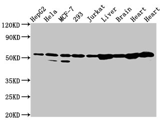
Western Blot Positive WB detected in: HepG2 whole cell lysate, Hela whole cell lysate, MCF-7 whole cell lysate, 293 whole cell lysate, Jurkat whole cell lysate, Rat liver tissue, Rat brain tissue, Rat heart tissue, Mouse heart tissue All lanes: DLD antibody at 2microg/ml Secondary Goat polyclonal to rabbit IgG at 1/50000 dilution Predicted band size: 55, 44, 50 kDa Observed band size: 55 kDa
. Section was blocked with 10% normal goat serum 30min at RT. Then primary antibody (1% BSA) was incubated at 4°C overnight. The primary is detected by a biotinylated secondary antibody and visualized using an HRP conjugated SP system.")
Western Blot Positive WB detected in: HepG2 whole cell lysate, Hela whole cell lysate, MCF-7 whole cell lysate, 293 whole cell lysate, Jurkat whole cell lysate, Rat liver tissue, Rat brain tissue, Rat heart tissue, Mouse heart tissue All lanes: DLD antibody at 2microg/ml Secondary Goat polyclonal to rabbit IgG at 1/50000 dilution Predicted band size: 55, 44, 50 kDa Observed band size: 55 kDa
DLD Antibody
CSB-PA006928LA01HU
ApplicationsWestern Blot, ELISA, ImmunoHistoChemistry
Product group Antibodies
ReactivityHuman, Mouse, Rat
TargetDLD
Overview
- SupplierCusabio
- Product NameDLD Antibody
- Delivery Days Customer20
- ApplicationsWestern Blot, ELISA, ImmunoHistoChemistry
- CertificationResearch Use Only
- ClonalityPolyclonal
- ConjugateUnconjugated
- Gene ID1738
- Target nameDLD
- Target descriptiondihydrolipoamide dehydrogenase
- Target synonymsDLDD, DLDH, E3, GCSL, LAD, OGDC-E3, PHE3, dihydrolipoyl dehydrogenase, mitochondrial, E3 component of pyruvate dehydrogenase complex, 2-oxo-glutarate complex, branched chain keto acid dehydrogenase complex, diaphorase, epididymis secretory sperm binding protein, glycine cleavage system L protein, glycine cleavage system protein L, lipoamide dehydrogenase, lipoamide reductase, lipoyl dehydrogenase, pyruvate dehydrogenase complex subunit E3, 2-oxo-glutarate complex, branched chain keto acid dehydrogenase complex, glycine cleavage system protein L
- HostRabbit
- IsotypeIgG
- Protein IDP09622
- Protein NameDihydrolipoyl dehydrogenase, mitochondrial
- Scientific DescriptionLipoamide dehydrogenase is a component of the glycine cleavage system as well as an E3 component of three alpha-ketoacid dehydrogenase complexes (pyruvate-, alpha-ketoglutarate-, and branched-chain amino acid-dehydrogenase complex). In monomeric form has additional moonlighting function as serine protease (PubMed:17404228). Involved in the hyperactivation of spermatazoa during capacitation and in the spermatazoal acrosome reaction (By similarity).
- ReactivityHuman, Mouse, Rat
- Storage Instruction-20°C or -80°C
- UNSPSC41116161
Related products

Product group Antibodies
Anti-DLD AntibodyA30484
ApplicationsImmunoFluorescence, Western Blot, ImmunoHistoChemistry
ReactivityHuman, Mouse, Rat
- SizePrice

Product group Antibodies
Anti-DLD AntibodyHPA044849
ApplicationsWestern Blot, ImmunoCytoChemistry, ImmunoHistoChemistry
ReactivityHuman
TargetDLD
- SizePrice
Product group Antibodies

DLD / Diaphorase / E3 AntibodyLS-C404893
ApplicationsELISA, ImmunoHistoChemistry
ReactivityHuman, Mouse, Rat
TargetDLD
- SizePrice
Product group Antibodies
DLD Polyclonal AntibodyCAC15094
ApplicationsWestern Blot, ELISA, ImmunoHistoChemistry
ReactivityMouse, Rat
TargetDLD
- SizePrice

Product group Antibodies
Anti-Lipoamide Dehydrogenase/DLD Antibody Picoband(r)PB9579-CARRIER-FREE
ApplicationsFlow Cytometry, ImmunoFluorescence, Western Blot, ImmunoCytoChemistry, ImmunoHistoChemistry
ReactivityBovine, Equine, Human, Monkey, Mouse, Rabbit, Rat
TargetDLD
- SizePrice
![Various whole cell extracts (30 μg) were separated by 10% SDS-PAGE, and the membrane was blotted with DLD antibody [N1N3] (GTX101232) diluted at 1:500. The HRP-conjugated anti-rabbit IgG antibody (GTX213110-01) was used to detect the primary antibody, and the signal was developed with Trident ECL plus-Enhanced.](https://www.genetex.com/upload/website/prouct_img/normal/GTX101232/GTX101232_40667_20181026_WB_R_w_23060100_154.webp)
Product group Antibodies
DLD antibody [N1N3]GTX101232
ApplicationsWestern Blot, ImmunoHistoChemistry, ImmunoHistoChemistry Paraffin
ReactivityHuman, Rat
TargetDLD
- SizePrice
Product group Antibodies
DLD Recombinant Antibody, AbBy Fluor-555 ConjugatedBSM-62296R-BF555
ApplicationsImmunoFluorescence, Western Blot
ReactivityHuman, Mouse, Rat
TargetDLD
- SizePrice
Product group Antibodies
Anti-DLD Antibody144-05403
ApplicationsWestern Blot, ImmunoHistoChemistry
ReactivityHuman, Mouse
TargetDLD
- SizePrice