

DOK2 antibody detects DOK2 protein by western blot analysis. Various whole cell extracts (30 μg) were separated by 10% SDS-PAGE, and the membrane was blotted with DOK2 antibody (GTX101577) diluted at a dilution of 1:10000.
![DOK2 antibody [N1C1] detects DOK2 protein at cytoplasm by immunofluorescent analysis. Sample: Jurkat cells were fixed in 4% paraformaldehyde at RT for 15 min. Green: DOK2 protein stained by DOK2 antibody [N1C1] (GTX101577) diluted at 1:500. Blue: Hoechst 33342 staining. Scale bar = 10 μm.](https://www.genetex.com/upload/website/prouct_img/normal/GTX101577/GTX101577_40618_IFA_w_23060100_139.webp "DOK2 antibody [N1C1] detects DOK2 protein at cytoplasm by immunofluorescent analysis. Sample: Jurkat cells were fixed in 4% paraformaldehyde at RT for 15 min. Green: DOK2 protein stained by DOK2 antibody [N1C1] (GTX101577) diluted at 1:500. Blue: Hoechst 33342 staining. Scale bar = 10 μm.")
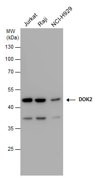
![DOK2 antibody [N1C1] detects DOK2 protein by western blot analysis. Whole cell extracts (30 μg) was separated by 10% SDS-PAGE, and the membrane was blotted with DOK2 antibody [N1C1] (GTX101577) at a dilution of 1:10000.](https://www.genetex.com/upload/website/prouct_img/normal/GTX101577/GTX101577_40618_WB_w_23060100_264.webp "DOK2 antibody [N1C1] detects DOK2 protein by western blot analysis. Whole cell extracts (30 μg) was separated by 10% SDS-PAGE, and the membrane was blotted with DOK2 antibody [N1C1] (GTX101577) at a dilution of 1:10000.")
DOK2 antibody detects DOK2 protein by western blot analysis. Various whole cell extracts (30 μg) were separated by 10% SDS-PAGE, and the membrane was blotted with DOK2 antibody (GTX101577) diluted at a dilution of 1:10000.
DOK2 antibody [N1C1]
GTX101577
ApplicationsImmunoFluorescence, Western Blot, ImmunoCytoChemistry
Product group Antibodies
ReactivityHuman
TargetDOK2
Overview
- SupplierGeneTex
- Product NameDOK2 antibody [N1C1]
- Delivery Days Customer9
- Application Supplier NoteWB: 1:5000-1:20000. ICC/IF: 1:100-1:1000. *Optimal dilutions/concentrations should be determined by the researcher.Not tested in other applications.
- ApplicationsImmunoFluorescence, Western Blot, ImmunoCytoChemistry
- CertificationResearch Use Only
- ClonalityPolyclonal
- Concentration1 mg/ml
- ConjugateUnconjugated
- Gene ID9046
- Target nameDOK2
- Target descriptiondocking protein 2
- Target synonymsp56DOK, p56dok-2, docking protein 2, docking protein 2, 56kD, docking protein 2, 56kDa, downstream of tyrosine kinase 2, p56(dok-2)
- HostRabbit
- IsotypeIgG
- Protein IDO60496
- Protein NameDocking protein 2
- Scientific DescriptionThe protein encoded by this gene is constitutively tyrosine phosphorylated in hematopoietic progenitors isolated from chronic myelogenous leukemia (CML) patients in the chronic phase. It may be a critical substrate for p210(bcr/abl), a chimeric protein whose presence is associated with CML. This encoded protein binds p120 (RasGAP) from CML cells. [provided by RefSeq]
- ReactivityHuman
- Storage Instruction-20°C or -80°C,2°C to 8°C
- UNSPSC41116161
Datasheet
Related products
Product group Antibodies
ApplicationsImmunoFluorescence, Western Blot, ImmunoHistoChemistry
ReactivityHuman, Mouse
- SizePrice
Product group Antibodies

DOK2 AntibodyLS-C749101
ApplicationsWestern Blot
ReactivityHuman, Mouse
TargetDOK2
- SizePrice
Product group Antibodies
DOK2 Polyclonal AntibodyBS-2746R
ApplicationsImmunoFluorescence, Western Blot, ELISA, ImmunoCytoChemistry, ImmunoHistoChemistry, ImmunoHistoChemistry Frozen, ImmunoHistoChemistry Paraffin
ReactivityBovine, Canine, Equine, Human, Mouse, Porcine, Rabbit, Rat
TargetDOK2
- SizePrice
Product group Antibodies
DOK2 AntibodyCSB-PA002181
ApplicationsImmunoFluorescence, Western Blot, ELISA, ImmunoHistoChemistry
ReactivityHuman, Mouse
TargetDOK2
- SizePrice
Product group Antibodies
DOK2 Polyclonal AntibodyCAC12851
ApplicationsImmunoFluorescence, Western Blot, ELISA, ImmunoHistoChemistry
ReactivityMouse
TargetDOK2
- SizePrice
Product group Antibodies
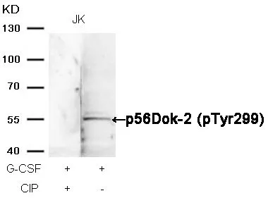
DOK2 (phospho Tyr299) antibodyGTX78982
ApplicationsWestern Blot, ImmunoHistoChemistry, ImmunoHistoChemistry Paraffin
ReactivityHuman
TargetDOK2
- SizePrice
Product group Antibodies
Anti-DOK2 AntibodyHPA005507
ApplicationsImmunoCytoChemistry
ReactivityHuman
TargetDOK2
- SizePrice
Product group Antibodies
DOK2 (phospho Tyr299) antibodyGTX50299
ApplicationsImmunoFluorescence, Western Blot, ImmunoCytoChemistry, ImmunoHistoChemistry, ImmunoHistoChemistry Paraffin
ReactivityHuman
TargetDOK2
- SizePrice