

Western Blot analysis of Jurkat cell and Human fetal kidney tissue using MKP-3 Polyclonal Antibody at dilution of 1:450
Western Blot analysis of Jurkat cell and Human fetal kidney tissue using MKP-3 Polyclonal Antibody at dilution of 1:450
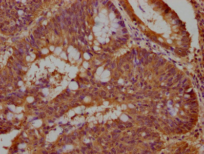
MKP-3 Polyclonal Antibody
E-AB-10308
ApplicationsWestern Blot
Product group Antibodies
TargetDUSP6
Overview
- SupplierElabscience
- Product NameMKP-3 Polyclonal Antibody
- Delivery Days Customer12
- ApplicationsWestern Blot
- Applications SupplierELISA WB IHC
- CertificationResearch Use Only
- ClonalityPolyclonal
- Concentration0.4 mg/ml
- ConjugateUnconjugated
- Gene ID1848
- Target nameDUSP6
- Target descriptiondual specificity phosphatase 6
- Target synonymsHH19, MKP3, PYST1, dual specificity protein phosphatase 6, MAP kinase phosphatase 3, dual specificity protein phosphatase PYST1, mitogen-activated protein kinase phosphatase 3, serine/threonine specific protein phosphatase
- HostRabbit
- IsotypeIgG
- Protein IDQ16828
- Protein NameDual specificity protein phosphatase 6
- Scientific DescriptionThe protein encoded by this gene is a member of the dual specificity protein phosphatase subfamily. These phosphatases inactivate their target kinases by dephosphorylating both the phosphoserine/threonine and phosphotyrosine residues. They negatively regulate members of the mitogen-activated protein (MAP) kinase superfamily (MAPK/ERK, SAPK/JNK, p38), which are associated with cellular proliferation and differentiation. Different members of the family of dual specificity phosphatases show distinct substrate specificities for various MAP kinases, different tissue distribution and subcellular localization, and different modes of inducibility of their expression by extracellular stimuli. This gene product inactivates ERK2, is expressed in a variety of tissues with the highest levels in heart and pancreas, and unlike most other members of this family, is localized in the cytoplasm. Two transcript variants encoding different isoforms have been found for this gene.
- Storage Instruction-20°C
- UNSPSC41116161
MSDS
Related products
Product group Antibodies

DCHS1 AntibodyLS-C154989
ApplicationsELISA
ReactivityHamster, Mouse
TargetDCHS1
- SizePrice

Product group Antibodies
Anti-DCHS1 AntibodyHPA050246
ApplicationsImmunoHistoChemistry
ReactivityHuman
TargetDCHS1
- SizePrice
Product group Antibodies
DCHS1 AntibodyCSB-PA850320OA01HU
ApplicationsImmunoFluorescence, ELISA, ImmunoHistoChemistry
ReactivityHuman
TargetDCHS1
- SizePrice
Product group Antibodies
DCHS1 AntibodyPACO63471
ApplicationsImmunoFluorescence, ELISA, ImmunoHistoChemistry
ReactivityHuman
TargetDCHS1
- SizePrice
Product group Antibodies
References
DUSP6 Polyclonal AntibodyBS-11546R
ApplicationsImmunoFluorescence, Western Blot, ImmunoHistoChemistry, ImmunoHistoChemistry Paraffin
ReactivityBovine, Canine, Equine, Human, Mouse, Rabbit, Rat
TargetDCHS1
- SizePrice