

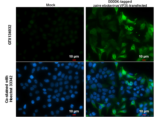
Ebola virus VP35 antibody detects Ebola virus VP35 protein at cytoplasm by immunofluorescent analysis. Sample: Mock and transfected 293T cells were fixed in 4% paraformaldehyde at RT for 15 min. Green: Ebola virus VP35 protein stained by Ebola virus VP35 antibody (GTX134032) diluted at 1:1000. Blue: Hoechst 33342 staining. Scale bar = 10 μm.
 and transfected (+) 293T whole cell extracts (5 μg) were separated by 10% SDS-PAGE, and the membranes were blotted with Ebola virus VP35 antibody (GTX134032) diluted at 1:5000 and DDDDK tag antibody (GTX115043) diluted at 1:2000. The HRP-conjugated anti-rabbit IgG antibody (GTX213110-01) was used to detect the primary antibody.")
Ebola virus VP35 antibody detects Ebola virus VP35 protein at cytoplasm by immunofluorescent analysis. Sample: Mock and transfected 293T cells were fixed in 4% paraformaldehyde at RT for 15 min. Green: Ebola virus VP35 protein stained by Ebola virus VP35 antibody (GTX134032) diluted at 1:1000. Blue: Hoechst 33342 staining. Scale bar = 10 μm.
Ebola virus VP35 antibody
GTX134032
ApplicationsImmunoFluorescence, Western Blot, ImmunoCytoChemistry
Product group Antibodies
ReactivityVirus
Overview
- SupplierGeneTex
- Product NameEbola virus VP35 antibody
- Delivery Days Customer9
- Application Supplier NoteWB: 1:1000-1:10000. ICC/IF: 1:100-1:1000. *Optimal dilutions/concentrations should be determined by the researcher.Not tested in other applications.
- ApplicationsImmunoFluorescence, Western Blot, ImmunoCytoChemistry
- CertificationResearch Use Only
- ClonalityPolyclonal
- Concentration0.55 mg/ml
- ConjugateUnconjugated
- HostRabbit
- IsotypeIgG
- ReactivityVirus
- Storage Instruction-20°C or -80°C,2°C to 8°C
- UNSPSC41116161