

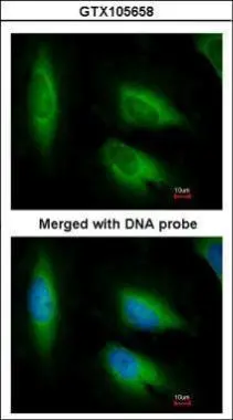
Immunofluorescence analysis of paraformaldehyde-fixed HeLa, using EEF1E1(GTX105658) antibody at 1:200 dilution.
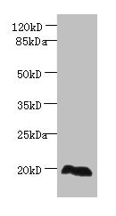
 A:A431(GTX27909) 12% SDS PAGE GTX105658 diluted at 1:1000")
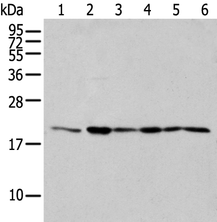
![Wild-type (WT) and EEF1E1 knockout (KO) 293T cell extracts (30 μg) were separated by 12% SDS-PAGE, and the membrane was blotted with EEF1E1 antibody [N1C3] (GTX105658) diluted at 1:500. The HRP-conjugated anti-rabbit IgG antibody (GTX213110-01) was used to detect the primary antibody.](https://www.genetex.com/upload/website/prouct_img/normal/GTX105658/GTX105658_39806_20180202_WB_KO_watermark_w_23060120_510.webp "Wild-type (WT) and EEF1E1 knockout (KO) 293T cell extracts (30 μg) were separated by 12% SDS-PAGE, and the membrane was blotted with EEF1E1 antibody [N1C3] (GTX105658) diluted at 1:500. The HRP-conjugated anti-rabbit IgG antibody (GTX213110-01) was used to detect the primary antibody.")
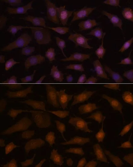
![EEF1E1 antibody [N1C3] detects EEF1E1 protein at cytoplasm on mouse fore brain by immunohistochemical analysis. Sample: Paraffin-embedded mouse fore brain. EEF1E1 antibody [N1C3] (GTX105658) diluted at 1:500.
Antigen Retrieval: Trilogy? (EDTA based, pH 8.0) buffer, 15min](https://www.genetex.com/upload/website/prouct_img/normal/GTX105658/GTX105658_39806_20141205_IHC_M_2_w_23060120_279.webp "EEF1E1 antibody [N1C3] detects EEF1E1 protein at cytoplasm on mouse fore brain by immunohistochemical analysis. Sample: Paraffin-embedded mouse fore brain. EEF1E1 antibody [N1C3] (GTX105658) diluted at 1:500.
Antigen Retrieval: Trilogy? (EDTA based, pH 8.0) buffer, 15min")
![EEF1E1 antibody [N1C3] detects EEF1E1 protein at cytoplasm on mouse fore brain by immunohistochemical analysis. Sample: Paraffin-embedded mouse fore brain. EEF1E1 antibody [N1C3] (GTX105658) diluted at 1:500.
Antigen Retrieval: Trilogy? (EDTA based, pH 8.0) buffer, 15min](https://www.genetex.com/upload/website/prouct_img/normal/GTX105658/GTX105658_39806_20141205_IHC_M_w_23060120_955.webp "EEF1E1 antibody [N1C3] detects EEF1E1 protein at cytoplasm on mouse fore brain by immunohistochemical analysis. Sample: Paraffin-embedded mouse fore brain. EEF1E1 antibody [N1C3] (GTX105658) diluted at 1:500.
Antigen Retrieval: Trilogy? (EDTA based, pH 8.0) buffer, 15min")
 A: Mouse brain 12% SDS PAGE GTX105658 diluted at 1:1000")
Immunofluorescence analysis of paraformaldehyde-fixed HeLa, using EEF1E1(GTX105658) antibody at 1:200 dilution.
EEF1E1 antibody [N1C3]
GTX105658
ApplicationsImmunoFluorescence, Western Blot, ImmunoCytoChemistry, ImmunoHistoChemistry, ImmunoHistoChemistry Paraffin
Product group Antibodies
ReactivityHuman, Mouse
TargetEEF1E1
Overview
- SupplierGeneTex
- Product NameEEF1E1 antibody [N1C3]
- Delivery Days Customer9
- Application Supplier NoteWB: 1:500-1:3000. ICC/IF: 1:100-1:1000. IHC-P: 1:100-1:1000. *Optimal dilutions/concentrations should be determined by the researcher.Not tested in other applications.
- ApplicationsImmunoFluorescence, Western Blot, ImmunoCytoChemistry, ImmunoHistoChemistry, ImmunoHistoChemistry Paraffin
- CertificationResearch Use Only
- ClonalityPolyclonal
- Concentration0.2 mg/ml
- ConjugateUnconjugated
- Gene ID9521
- Target nameEEF1E1
- Target descriptioneukaryotic translation elongation factor 1 epsilon 1
- Target synonymsAIMP3, P18, eukaryotic translation elongation factor 1 epsilon-1, ARS-interacting multifunctional protein 3, aminoacyl tRNA synthetase complex-interacting multifunctional protein 3, multisynthase complex auxiliary component p18, multisynthetase complex auxiliary component p18, p18 component of aminoacyl-tRNA synthetase complex
- HostRabbit
- IsotypeIgG
- Protein IDO43324
- Protein NameEukaryotic translation elongation factor 1 epsilon-1
- Scientific DescriptionThis gene encodes a multifunctional protein that localizes both in the cytoplasm and in the nucleus. In the cytoplasm, the encoded protein is an auxiliary component of the macromolecular aminoacyl-tRNA synthase complex. However, its mouse homolog has been shown to translocate to the nucleus in response to DNA damage and plays a positive role in ATM/ATR-mediated p53 activation. Alternative splicing results in multiple transcript variants. [provided by RefSeq]
- ReactivityHuman, Mouse
- Storage Instruction-20°C or -80°C,2°C to 8°C
- UNSPSC41116161
Datasheet
Related products
Product group Antibodies
EEF1E1 AntibodyCSB-PA007432LA01HU
ApplicationsImmunoFluorescence, Western Blot, ELISA, ImmunoHistoChemistry
ReactivityHuman
TargetEEF1E1
- SizePrice
Product group Antibodies
Anti-EEF1E1 AntibodyA37392
ApplicationsWestern Blot, ImmunoHistoChemistry
ReactivityHuman
- SizePrice

Product group Antibodies
Anti-EEF1E1 AntibodyM08532
ApplicationsFlow Cytometry, ImmunoFluorescence, Western Blot
ReactivityHuman, Mouse
TargetEEF1E1
- SizePrice
Product group Antibodies

EEF1E1 / AIMP3 AntibodyLS-C830353
ApplicationsWestern Blot, ELISA, ImmunoHistoChemistry
ReactivityHuman, Mouse
TargetEEF1E1
- SizePrice

Product group Antibodies
Anti-EEF1E1 AntibodyHPA027901
ApplicationsWestern Blot, ImmunoHistoChemistry
ReactivityHuman
TargetEEF1E1
- SizePrice
Product group Antibodies

ApplicationsWestern Blot, ELISA, ImmunoHistoChemistry
ReactivityBovine, Canine, Human, Porcine
TargetEEF1E1
- SizePrice
Product group Antibodies
Eef1E1 Polyclonal AntibodyCAC08384
ApplicationsImmunoFluorescence, Western Blot, ELISA, ImmunoHistoChemistry
TargetEEF1E1
- SizePrice
Product group Antibodies
EEF1E1 antibodyGTX66367
ApplicationsImmunoFluorescence, Western Blot, ImmunoCytoChemistry
ReactivityHuman, Mouse, Rat
TargetEEF1E1
- SizePrice
Product group Antibodies
Anti-EEF1E1Y158129
ApplicationsWestern Blot, ELISA, ImmunoHistoChemistry
ReactivityHuman, Mouse, Rat
- SizePrice