

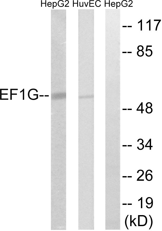
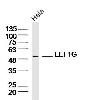
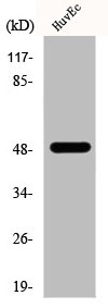
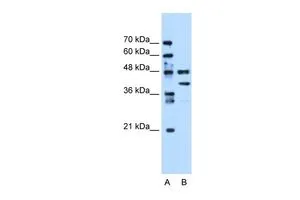
Various whole cell extracts (30 μg) were separated by 10% SDS-PAGE, and the membrane was blotted with EEF1G antibody [GT1278] (GTX03190) diluted at 1:1000. The HRP-conjugated anti-rabbit IgG antibody (GTX213110-01) was used to detect the primary antibody.
![ICC/IF analysis of NIH-3T3 cells using GTX03190 EEF1G antibody [GT1278]. Blue : DAPI for nuclear staining Dilution : 1:100](https://www.genetex.com/upload/website/prouct_img/normal/GTX03190/GTX03190_20210615_ICCIF_44_w_23053123_749.webp "ICC/IF analysis of NIH-3T3 cells using GTX03190 EEF1G antibody [GT1278]. Blue : DAPI for nuclear staining Dilution : 1:100")
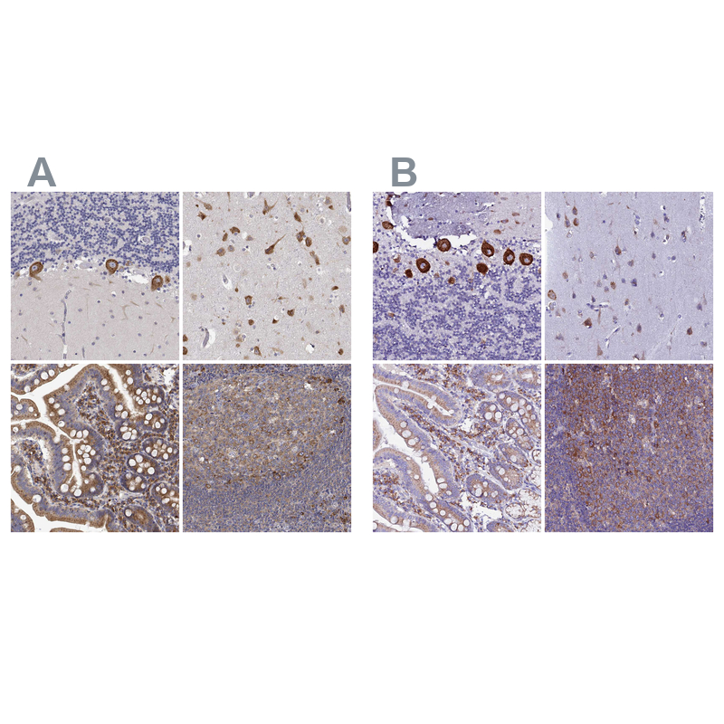
![IHC-P analysis of rat brain tissue section using GTX03190 EEF1G antibody [GT1278]. Dilution : 1:100](https://www.genetex.com/upload/website/prouct_img/normal/GTX03190/GTX03190_20210615_IHC-P_40_w_23053123_820.webp "IHC-P analysis of rat brain tissue section using GTX03190 EEF1G antibody [GT1278]. Dilution : 1:100")
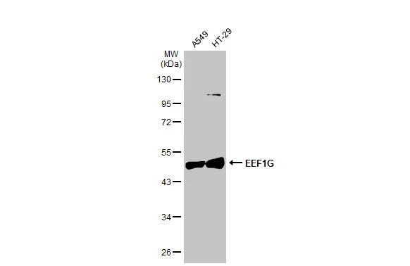
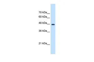
![WB analysis of various samples using GTX03190 EEF1G antibody [GT1278]. Dilution : 1:1000 Loading : 25μg per lane](https://www.genetex.com/upload/website/prouct_img/normal/GTX03190/GTX03190_47_WB_w_23053123_953.webp "WB analysis of various samples using GTX03190 EEF1G antibody [GT1278]. Dilution : 1:1000 Loading : 25μg per lane")
![ICC/IF analysis of HeLa cells using GTX03190 EEF1G antibody [GT1278]. Blue : DAPI for nuclear staining Dilution : 1:100](https://www.genetex.com/upload/website/prouct_img/normal/GTX03190/GTX03190_20210615_ICCIF_43_w_23053123_998.webp "ICC/IF analysis of HeLa cells using GTX03190 EEF1G antibody [GT1278]. Blue : DAPI for nuclear staining Dilution : 1:100")
![IHC-P analysis of mouse brain tissue section using GTX03190 EEF1G antibody [GT1278]. Dilution : 1:100](https://www.genetex.com/upload/website/prouct_img/normal/GTX03190/GTX03190_20210615_IHC-P_42_w_23053123_968.webp "IHC-P analysis of mouse brain tissue section using GTX03190 EEF1G antibody [GT1278]. Dilution : 1:100")
![Various whole cell extracts (30 μg) were separated by 10% SDS-PAGE, and the membrane was blotted with EEF1G antibody [GT1278] (GTX03190) diluted at 1:1000. The HRP-conjugated anti-rabbit IgG antibody (GTX213110-01) was used to detect the primary antibody.](https://www.genetex.com/upload/website/prouct_img/normal/GTX03190/GTX03190_4000001287_20210625_WB_w_23053123_501.webp "Various whole cell extracts (30 μg) were separated by 10% SDS-PAGE, and the membrane was blotted with EEF1G antibody [GT1278] (GTX03190) diluted at 1:1000. The HRP-conjugated anti-rabbit IgG antibody (GTX213110-01) was used to detect the primary antibody.")
![IHC-P analysis of human placenta tissue section using GTX03190 EEF1G antibody [GT1278]. Dilution : 1:100](https://www.genetex.com/upload/website/prouct_img/normal/GTX03190/GTX03190_20210615_IHC-P_41_w_23053123_267.webp "IHC-P analysis of human placenta tissue section using GTX03190 EEF1G antibody [GT1278]. Dilution : 1:100")
Various whole cell extracts (30 μg) were separated by 10% SDS-PAGE, and the membrane was blotted with EEF1G antibody [GT1278] (GTX03190) diluted at 1:1000. The HRP-conjugated anti-rabbit IgG antibody (GTX213110-01) was used to detect the primary antibody.
EEF1G antibody [GT1278]
GTX03190
ApplicationsImmunoFluorescence, Western Blot, ImmunoCytoChemistry, ImmunoHistoChemistry, ImmunoHistoChemistry Paraffin
Product group Antibodies
ReactivityHuman, Mouse, Rat
TargetEEF1G
Overview
- SupplierGeneTex
- Product NameEEF1G antibody [GT1278]
- Delivery Days Customer9
- Application Supplier NoteWB: 1:500 - 1:2000. ICC/IF: 1:50 - 1:200. IHC-P: 1:50 - 1:200. *Optimal dilutions/concentrations should be determined by the researcher.Not tested in other applications.
- ApplicationsImmunoFluorescence, Western Blot, ImmunoCytoChemistry, ImmunoHistoChemistry, ImmunoHistoChemistry Paraffin
- CertificationResearch Use Only
- ClonalityMonoclonal
- Clone IDGT1278
- Concentration0.32 mg/ml
- ConjugateUnconjugated
- Gene ID1937
- Target nameEEF1G
- Target descriptioneukaryotic translation elongation factor 1 gamma
- Target synonymsEF1G, GIG35, elongation factor 1-gamma, EF-1-gamma, PRO1608, eEF-1B gamma, pancreatic tumor-related protein, translation elongation factor eEF-1 gamma chain
- HostRabbit
- IsotypeIgG
- Protein IDP26641
- Protein NameElongation factor 1-gamma
- Scientific DescriptionThis gene encodes a subunit of the elongation factor-1 complex, which is responsible for the enzymatic delivery of aminoacyl tRNAs to the ribosome. This subunit contains an N-terminal glutathione transferase domain, which may be involved in regulating the assembly of multisubunit complexes containing this elongation factor and aminoacyl-tRNA synthetases. [provided by RefSeq, Jul 2008]
- ReactivityHuman, Mouse, Rat
- Storage Instruction-20°C or -80°C,2°C to 8°C
- UNSPSC41116161
Datasheet
Related products
Product group Antibodies
Anti-EEF1G AntibodyA97575
ApplicationsWestern Blot, ELISA
ReactivityHuman, Mouse, Rat
- SizePrice
Product group Antibodies
Anti-EEF1G Antibody144-07891
ApplicationsWestern Blot, ImmunoHistoChemistry
ReactivityHuman, Mouse, Rat
TargetEEF1G
- SizePrice

Product group Antibodies
ApplicationsFlow Cytometry, ImmunoFluorescence, ImmunoPrecipitation, Western Blot, ImmunoCytoChemistry, ImmunoHistoChemistry
ReactivityHuman, Mouse, Rat
TargetEEF1G
- SizePrice
Product group Antibodies
References
EEF1G Polyclonal AntibodyBS-13056R
ApplicationsImmunoFluorescence, Western Blot, ImmunoCytoChemistry, ImmunoHistoChemistry, ImmunoHistoChemistry Frozen, ImmunoHistoChemistry Paraffin
ReactivityBovine, Canine, Equine, Human, Mouse, Porcine, Rabbit, Rat
TargetEEF1G
- SizePrice
Product group Antibodies
EEF1G AntibodyCSB-PA002259
ApplicationsWestern Blot, ELISA
ReactivityHuman, Mouse, Rat
TargetEEF1G
- SizePrice
Product group Antibodies
ApplicationsImmunoPrecipitation, Western Blot, ImmunoCytoChemistry, ImmunoHistoChemistry
ReactivityMouse, Rat
TargetEEF1G
- SizePrice
Product group Antibodies

EF1G / EEF1G AntibodyLS-C403973
ApplicationsWestern Blot, ELISA, ImmunoHistoChemistry
ReactivityHuman, Mouse, Rat
TargetEEF1G
- SizePrice
Product group Antibodies
Anti-EEF1G AntibodyHPA055316
ApplicationsImmunoHistoChemistry
ReactivityHuman
TargetEEF1G
- SizePrice
Product group Antibodies
EEF1G antibody, InternalGTX46253
ApplicationsWestern Blot
ReactivityHuman
TargetEEF1G
- SizePrice
Product group Antibodies
EEF1G antibody, N-termGTX46254
ApplicationsWestern Blot
ReactivityHuman
TargetEEF1G
- SizePrice