

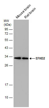
Various tissue extracts (50 μg) were separated by 12% SDS-PAGE, and the membrane was blotted with EFHD2 antibody (GTX108080) diluted at 1:1000.
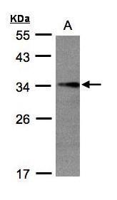
 and transfected (+) 293T whole cell extracts (30 μg) were separated by 12% SDS-PAGE, and the membrane was blotted with EFHD2 antibody (GTX108080) diluted at 1:1000. The HRP-conjugated anti-rabbit IgG antibody (GTX213110-01) was used to detect the primary antibody.")
![EFHD2 antibody detects EFHD2 protein expression by immunohistochemical analysis. Sample: Frozen sectioned E13.5 Rat brain. Green: EFHD2 protein stained by EFHD2 antibody (GTX108080) diluted at 1:250. Red: beta Tubulin 3/ TUJ1, a mature neuron marker, stained by beta Tubulin 3/ TUJ1 antibody [GT11710] (GTX631836) diluted at 1:500. Blue: Fluoroshield with DAPI (GTX30920).](https://www.genetex.com/upload/website/prouct_img/normal/GTX108080/GTX108080_39799_20160921_IHC-Fr_w_23060120_183.webp "EFHD2 antibody detects EFHD2 protein expression by immunohistochemical analysis. Sample: Frozen sectioned E13.5 Rat brain. Green: EFHD2 protein stained by EFHD2 antibody (GTX108080) diluted at 1:250. Red: beta Tubulin 3/ TUJ1, a mature neuron marker, stained by beta Tubulin 3/ TUJ1 antibody [GT11710] (GTX631836) diluted at 1:500. Blue: Fluoroshield with DAPI (GTX30920).")
![EFHD2 antibody detects EFHD2 protein by immunofluorescent analysis. Sample: DIV14 rat E18 primary cortical neurons were fixed in 4% paraformaldehyde at RT for 15 min. Green: EFHD2 protein stained by EFHD2 antibody (GTX108080) diluted at 1:500. Red: beta Tubulin 3/ Tuj1, stained by beta Tubulin 3/ Tuj1 antibody [GT1338] (GTX631831) diluted at 1:500. Blue: Fluoroshield with DAPI (GTX30920).](https://www.genetex.com/upload/website/prouct_img/normal/GTX108080/GTX108080_39799_20170719_IFA_R_w_23060120_886.webp "EFHD2 antibody detects EFHD2 protein by immunofluorescent analysis. Sample: DIV14 rat E18 primary cortical neurons were fixed in 4% paraformaldehyde at RT for 15 min. Green: EFHD2 protein stained by EFHD2 antibody (GTX108080) diluted at 1:500. Red: beta Tubulin 3/ Tuj1, stained by beta Tubulin 3/ Tuj1 antibody [GT1338] (GTX631831) diluted at 1:500. Blue: Fluoroshield with DAPI (GTX30920).")
 A:H1299 10% SDS PAGE GTX108080 diluted at 1:1000")
Various tissue extracts (50 μg) were separated by 12% SDS-PAGE, and the membrane was blotted with EFHD2 antibody (GTX108080) diluted at 1:1000.
EFHD2 antibody
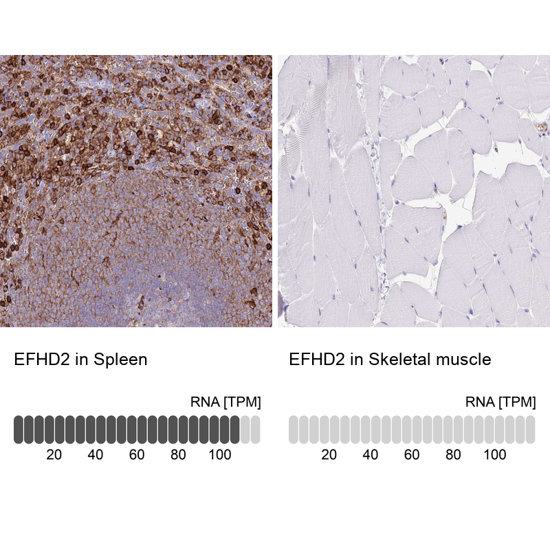
GTX108080
ApplicationsImmunoFluorescence, Western Blot, ImmunoCytoChemistry, ImmunoHistoChemistry, ImmunoHistoChemistry Frozen
Product group Antibodies
ReactivityHuman, Mouse, Rat
TargetEFHD2
Overview
- SupplierGeneTex
- Product NameEFHD2 antibody
- Delivery Days Customer9
- Application Supplier NoteWB: 1:500-1:3000. ICC/IF: 1:100-1:1000. IHC-Fr: 1:100-1:1000. *Optimal dilutions/concentrations should be determined by the researcher.Not tested in other applications.
- ApplicationsImmunoFluorescence, Western Blot, ImmunoCytoChemistry, ImmunoHistoChemistry, ImmunoHistoChemistry Frozen
- CertificationResearch Use Only
- ClonalityPolyclonal
- Concentration0.2 mg/ml
- ConjugateUnconjugated
- Gene ID79180
- Target nameEFHD2
- Target descriptionEF-hand domain family member D2
- Target synonymsSWS1, EF-hand domain-containing protein D2, EF hand domain containing 2, swiprosin 1, testicular tissue protein Li 62
- HostRabbit
- IsotypeIgG
- Protein IDQ96C19
- Protein NameEF-hand domain-containing protein D2
- Scientific DescriptionMay regulate B-cell receptor (BCR)-induced immature and primary B-cell apoptosis (By similarity). Plays a role as negative regulator of the canonical NF-kappa-B-activating branch (By similarity). Controls spontaneous apoptosis through the regulation of BCL2L1 abundance.
- ReactivityHuman, Mouse, Rat
- Storage Instruction-20°C or -80°C,2°C to 8°C
- UNSPSC41116161
Datasheet
Related products

Product group Antibodies
Anti-EFHD2 AntibodyA07124
ApplicationsImmunoFluorescence, Western Blot, ELISA, ImmunoHistoChemistry, ImmunoHistoChemistry Paraffin
ReactivityHuman, Mouse, Rat
TargetEFHD2
- SizePrice
Product group Antibodies
Anti-EFHD2 Antibody144-60853
ApplicationsWestern Blot
ReactivityHuman, Mouse
TargetEFHD2
- SizePrice
Product group Antibodies

EFHD2 AntibodyLS-C749906
ApplicationsWestern Blot
ReactivityHuman, Mouse
TargetEFHD2
- SizePrice
Product group Antibodies
EFHD2 Polyclonal AntibodyBS-14520R
ApplicationsImmunoFluorescence, Western Blot, ELISA, ImmunoCytoChemistry, ImmunoHistoChemistry, ImmunoHistoChemistry Frozen, ImmunoHistoChemistry Paraffin
ReactivityBovine, Human, Mouse, Porcine, Rat, Sheep
TargetEFHD2
- SizePrice
Product group Antibodies
EFHD2 AntibodyCSB-PA856907LA01HU
ApplicationsImmunoFluorescence, ELISA, ImmunoHistoChemistry
ReactivityHuman
TargetEFHD2
- SizePrice
Product group Antibodies
EFHD2 antibody, C-termGTX89662
ApplicationsWestern Blot, ImmunoHistoChemistry, ImmunoHistoChemistry Paraffin
ReactivityHuman
TargetEFHD2
- SizePrice
Product group Antibodies
Anti-EFHD2 AntibodyHPA048961
ApplicationsWestern Blot, ImmunoCytoChemistry, ImmunoHistoChemistry
ReactivityHuman
TargetEFHD2
- SizePrice